Upgrade to Pro — share decks privately, control downloads, hide ads and more …

Cryotherapy_Powerpoint_Updated_May_2025.pdf

 Cryotherapy_Powerpoint_Updated_May_2025.pdf

Transcript

  1. Course Overview • 1. Aims & Objectives • 2. History

    of Cryotherapy • 3. Cryotherapy Science & Mechanisms • 4. Dermatological Lesions • 5. CryoPen Use & Health & Safety • 6. Treatment Protocols • 7. Contraindications & Contra-actions • 8. Client Consultation & Aftercare
  2. Aims and Objectives The course aims to ensure you; the

    student understands the basics of health and safety and anatomy and physiology of the treatment. This manual covers the treatment background, benefits, consultation and contra-indications, contra-actions, aftercare and equipment and products required to perform the treatment. The practical techniques will be covered on the practical session to ensure competency in the procedure. At the end of the course, you will be able to perform a treatment in a professional, safe and hygienic manner in a commercially acceptable time, along with experience in carrying out a thorough consultation with the knowledge of the background, benefits, consultation, contra- indications, contra-actions, aftercare, equipment and the products needed.
  3. *Medical Exclaimer* It is advised that you take medical advice

    if you or any of your clients have a health problem. Any qualification from (Beautiful World Training Academy) will not qualify you to advise on or diagnose any medical condition.
  4. Ancient Beginnings • Used as an analgesic and anti-inflammatory by

    Egyptians and Greeks. • Hippocrates (5th century BC) applied cold to reduce bleeding and swelling. 19th Century Innovation • Dr. James Arnott (Scotland) pioneered medical freezing (~−20°C). • Used ice-salt mixtures to treat tumours and inflammation. • Preferred cold over chemical anaesthesia due to fewer side effects. The Cold Race Begins • Search for colder, controlled refrigerants led to the liquefaction of gases. • Enabled precise cryogenic tools for medical use. From Ice Packs to Precision • Solid CO₂, liquid air, and later liquid nitrogen became cryo agents. • Devices evolved from crude flasks to handheld CryoPens. Modern Cryotherapy • Now used across dermatology, oncology, gynaecology and more. • Combines ancient wisdom with cutting-edge science.
  5. Cryotherapy What is Cryotherapy? Controlled Cold for Therapeutic Effect Cryotherapy

    involves applying cold temperatures to the skin or tissue to induce a therapeutic response — without causing permanent damage. How It Works • Freeze–Thaw Cycles cause cell dehydration, ice crystal formation, and vascular stasis. • Results in reduced inflammation, pain relief, and targeted lesion destruction. Key Mechanisms • Boiling Transfer: Direct cryogen spray (e.g. liquid nitrogen). • Conduction Transfer: CryoPens. • Cell Death: Ranges from reversible (apoptosis) to irreversible (necrosis) based on temperature and exposure. Common Cryogens Used • Liquid Nitrogen (−196°C) • Nitrous Oxide (−89°C) • Carbon Dioxide (−78.5°C) • Used For: Skin tags, warts, solar lentigines, cherry angiomas, seborrhoeic keratoses & more.
  6. Primary Lesion Primary skin lesions are variations in colour or

    texture that may be present at birth, such as moles or birthmarks or something that appears over time such as those associated with infectious diseases, for example; warts, acne or psoriasis, allergic reactions such as hives or contact dermatitis or environmental causes like sunburn. Macule: Flat, discoloured spot <1 cm Papule: Raised bump <1 cm, no fluid Nodule: Firm lump >1 cm, deeper in skin Vesicle: Small fluid-filled blister <1 cm Bullae: Large blister >1 cm Pustule: Pus-filled bump Wheal: Raised, itchy welt Urticaria: Hives caused by allergic reaction
  7. Secondary Lesion A secondary skin lesion is those changes in

    the skin that result from primary skin lesions, either as a natural progression or as a result of a person manipulating a primary lesion, for example, scratching or picking. Crust/Scab: Dried blood, serum or pus Scale: Flaky build-up of dead skin Excoriation: Scratch-induced damage Fissure: Linear skin crack Ulcer: Deep tissue loss, often inflamed Keloid: Overgrown scar tissue Scar: Healed fibrous tissue Moles: Pigmented lesions (monitor for changes)
  8. The CryoPen is primarily used for treating benign skin lesions

    — both vascular and non-vascular — that are clearly defined and superficial. Here's a list of common dermatological conditions suitable for CryoPen treatment: CryoPen is Used For: Vascular Lesions: Cherry Angiomas / Campbell de Morgan Spots Haemangiomas (superficial types) Spider Naevi (select cases)
  9. Non-Vascular Lesions: Skin Tags Solar Lentigines / Age Spots Seborrhoeic

    Keratoses Common Warts (including plantar and periungual) Milia Actinic Keratoses (only when confirmed non-malignant) CryoPen Should Not Be Used On: Suspicious moles or changing pigmented lesions Malignant skin cancers (e.g. melanoma, SCC, BCC) Lesions of unclear diagnosis without GP referral
  10. Macule Distinct discoloration of the skin, flat and smaller than

    1 cm in diameter. It does not cause a change in skin texture or thickness. Macules are noticed visually.
  11. Papule A raised skin area with no visible fluid and

    sized up to 1 cm in diameter. They have distinct borders and come in a variety of shapes.
  12. Nodule An elevated bump on the skin that is larger

    than a papule (greater than 1 cm in diameter). It may occur in all layers of the skin including the epidermis, dermis and subcutaneous tissue.
  13. Tumour A solid mass on the skin or subcutaneous tissue

    (under the skin). It is firm and usually larger than 2 cms.
  14. Vesicle A raised bump less than 1 cm in diameter

    and filled with air or clear liquid.
  15. Bullae Bullae are fluid-filled sacs that form when fluid is

    trapped under a thin layer of skin. They are similar to blisters and vesicles except that bullae have a diameter larger than 1 centimetre.
  16. Pustule A small bump on the skin containing pus. They

    can be infected but not always, as in the case of pustular psoriasis.
  17. Wheal A red, swollen mark that is often itchy and

    changes shape. They can occur in response to a stimulus such as a bug bite or food allergies. They are also known as welts or hives.
  18. Urticaria Also known as hives, welts or nettle rash and

    caused by an allergen. This results in widespread itchy skin – not just on the area that has been directly affected.
  19. Ephelides Medical term for freckles. The singular form of the

    word is ephelis. Consists of dark spots on the skin comprising of the protein melanin.
  20. Port Wine Stain Port-wine stains are (present at birth) capillary

    malformations. The colour of the birthmark is either pink, red, or purple. As the child grows, the mark may become darker. Port-wine stains can appear on any part of the body and vary in size.
  21. Strawberry Birthmark A haemangioma, a collection of blood vessels that

    form a lump under the skin; often called a ‘strawberry mark’ as it looks like the surface of a strawberry. Superficial haemangiomas are usually raised, red areas of skin, which feel warm because the blood vessels are close to the surface. Initially appear as a small area of pale skin where a red spot develops. Deep haemangiomas are blue in colour because the blood vessels are deeper in the skin. You can’t usually see them for the first few weeks as they only show a lump as the haemangioma grows.
  22. Melasma/Chloasma Dark brown or grey discoloration of the skin that

    typically affects sun-exposed parts of the face. The greater the melanin is in skin, the more likely it is to experience melasma—this means those with darker skin tones are more likely to have it. Certain health conditions can also make skin more sensitive to sunlight and more prone to developing melasma, such as thyroid problems and being on birth control pills. It's also thought to be hereditary. Pregnancy can increase the risk as well. Melasma is referred to as the “mask of pregnancy” and can occur in up to 50% of pregnant women.
  23. Leucoderma/Vitiligo Vitiligo is a disease that causes the loss of

    skin colour in blotches. Extent and rate of pigment loss from vitiligo is unpredictable. Affects the skin on any part of the body. It can also affect hair and the inside of the mouth. The colour of hair and skin is determined by melanin. Vitiligo occurs when the cells that produce melanin stop functioning. Vitiligo affects people of all skin types, but it may be more noticeable in people with darker skin. Leucoderma is a skin disorder in which patches of skin tend to lose its natural colour. Regarded as the de- pigmentation of the skin which is seen by the localisation or complete destruction of melanocytes in the body. The characteristic formation of white patches on the skin remains closely bound to each other unlike the patches formed in Vitiligo. Localised to smaller areas in the beginning over time, the skin patches might get enlarged. Most of the major skin changes might become noticeable around the age of 10 to 30 years. Skin colour changes are more noticeable in people with darker skin complexions.
  24. Solar Lentignes / Liver Harmless patch of darkened skin. Caused

    from exposure to ultraviolet (UV) radiation, which causes local proliferation of melanocytes and accumulation of melanin within the skin cells (keratinocytes). Very common, especially in people over the age of 40 years. Also known as an “old age spot” or “senile freckle”. Flat, well-circumscribed patches can be round, oval or irregular in shape. Colour varies from skin-coloured, tan to dark brown or black, and size varies from a few mms to several cms in diameter. They can be slightly scaly. Found as groups of similar lesions on sun-exposed sites, particularly the face or the back of hands. They occur in light and dark skin but tend to be more numerous in fair-skinned individuals.
  25. Xanthelasma /Xanthoma Yellow flat plaques over the upper or lower

    eyelids, most often near the inner canthus. They represent areas of lipid-containing macrophages, but the exact pathophysiology is not known. In other areas of the body the individual lesion would be called a xanthoma; xanthelasma is the most common xanthoma.
  26. Scales A dry, horny build-up of dead skin cells that

    often flakes off the surface of the skin. Diseases that promote scale include fungal infections, psoriasis and seborrheic dermatitis
  27. Crust/Scab Dried collection of blood, serum or pus also called

    a scab. Often part of the normal healing process of many infections’ lesions.
  28. Fissure A cutaneous condition in which there is a linear-like

    cleavage of skin, sometimes defined as extending into the dermis. It is smaller than a skin laceration.
  29. Ulcer A sore on the skin or a mucous membrane,

    accompanied by the disintegration of tissue. Can result in complete loss of the epidermis and often portions of the dermis and even subcutaneous fat. Most common on the skin of the lower extremities and in the gastrointestinal tract. An ulcer that appears on the skin is often visible as an inflamed tissue with an area of reddened skin. Often visible in the event of exposure to heat or cold, irritation, or a problem with blood circulation. Can also be caused due to a lack of mobility, which causes prolonged pressure on the tissues. This stress in the blood circulation is transformed to a skin ulcer, commonly known as bedsores or decubitus ulcers. Ulcers often become infected, and pus forms.
  30. Keloid When skin is injured, fibrous tissue called scar tissue

    forms over the wound to repair and protect the injury. In some cases, scar tissue grows excessively, forming smooth, hard growths called keloids. These can be much larger than the original wound. They’re most commonly found on the chest, shoulders, earlobes, and cheeks. However, can affect any part of the body.
  31. Moles Normal Moles/Pigmented Naevi Common hyperpigmented skin lesions can vary

    in colour, size, shape and location. They are benign and are of no concern. They develop during the client’s life, and sun exposure increases the number of moles present. As beauty therapists, we are unable to treat moles, and we should refer clients to other professionals. Why moles occur is still being researched. Many types of moles can be hereditary, indicating a genetic predisposition. Fairer skin types generally have more moles, as do those who spend a lot of time in the sun. Moles can also change or appear due to hormonal changes such as puberty or pregnancy.
  32. Moles The characteristics of a normal mole will be: ⮚SYMMETRY

    – Symmetrical, round or oval ⮚BORDER - Well defined ⮚COLOUR - Flesh coloured, light brown or dark brown ⮚DIAMETER - Usually less than ½ cm ⮚EVOLUTION - Stay fairly static with little change throughout the client’s life ⮚LOCATION - Most commonly found on sun-exposed areas ⮚UNIFORMITY - Most moles look similar
  33. Common Moles Description: ⮚Have even pigmentation and a dome-like smooth

    surface with distinct edges. Typically found on skin that’s exposed to sun regularly and can, although very rarely turn into skin cancer.
  34. Junctional Melanocytic Naevi Description: These develop due to an accumulation

    of melanocytes that are located predominantly at the dermo-epidermal junction, hence their name. Often quite darkly pigmented and are macular or very thinly popular with only minimal elevation above the level of the skin. An acquired lesion and as they age change their characteristics to that of a compound naevus, where there are accumulations of melanocytes in the dermis and at the dermo-epidermal junction, which can cause the lesion to become increasingly popular. Can occur at any site on the body and are regularly shaped, usually round or oval. Normally uniform in colour and range in pigmentation from light to dark brown. They are usually under 7mm in diameter. They are benign lesions but may turn into melanoma.
  35. Dermal Melanocytic Naevi Description: This mole is a form of

    melanocytic naevus, appears to have the same degree of pigmentation as the surrounding skin. Raised from the skin similar to that of a classic mole. Melanocytes do not impart their pigmentation in the lesion as they are located deep within the dermis rather than the dermo-epidermal junction. May appear spontaneously or grow from a pre-existing pigmented mole. Usually develop from the end of childhood onwards and can appear at any stage throughout adulthood. They are small between 5mm-1cm, raised from the surface of the skin and may be associated with hair growth.
  36. Dysplastic Naevi Description: A dysplastic nevus is also known as

    an atypical mole. Its appearance is different from that of a common mole. Often grow larger than ordinary moles and may have irregular and indistinct borders. The colour may not be uniform and may range from light pink to very dark brown. Usually begin flat, but parts may raise above the skins surface over time. May be located anywhere but are more commonly found on the trunk on men and on calves of women.
  37. Halo Naevi Description: A benign skin lesion that is a

    result of a common melanocytic naevus undergoing an inflammatory process, such that a zone of depigmentation surrounds the mole. There is an infiltration of T lymphocytes and macrophages and possibly some anti-body – mediated autoimmunity. The aetiology and pathophysiology of the immune reaction to the presence of an aggregate of melanocytes is poorly understood.
  38. Congenital Naevi Description: A congenital mole is something that’s present

    at birth. Melanocyte cells in the dermis and epidermis can cause these types of moles. They can range in size and might be confused with birthmarks. They pose a risk of turning into melanoma.
  39. Mole Changes and Abnormalities If a mole changes any of

    its characteristics, it is essential that the client seeks the advice of a GP as malignant melanomas often develop from moles that the client may have had for many years. When a mole that becomes asymmetrical changing from oval or round to having an undefined irregular border or changes in size or colour, or starts to become itchy or bleed, then the client should have this checked immediately. Melanoma is caused mainly by intense, occasional UV exposure that may have frequently led to sunburn, especially in those who are genetically predisposed to the disease. Around 1 in 10 of people with melanoma will have family members that have also had one. There are many reasons for this. Fair skin is inherited; dysplastic naevi can run in families, as can a tendency to have a large number of ordinary moles present. Melanomas might not cause any symptoms at all, but a tingling or itching sensation might occur at an early stage. Minor changes in the size, shape or colour of an existing mole may occur, whilst others begin as a dark area that can look like a new mole. After a while, a melanoma may feel hard and lumpy, may bleed, ooze or crust up. Melanomas do not all look the same, and there are several different types. The ABCD system tells you some of the things to look out for.
  40. Melanoma A melanoma may show one or more of the

    following features: ⮚ASYMMETRY – the two halves of the area differ in their shape. ⮚BORDER – the edges of the area may be irregular or blurred and sometimes show notches. ⮚COLOUR – this may be uneven. Different shades of black, brown and pink may be seen. ⮚DIAMETER - most melanomas are at least 6 mm. in diameter. Melanomas can be present on any part of the body, but they are more common in men on the body and in women on their legs.
  41. Squamous Cell Carcinoma The most second form of skin cancer

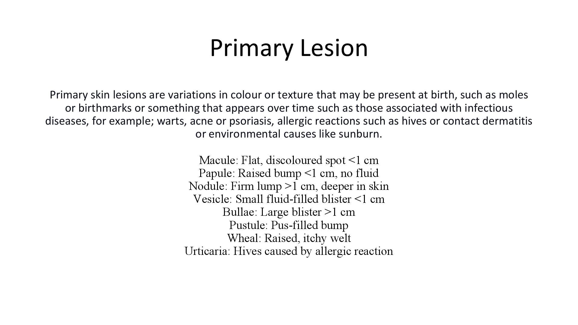
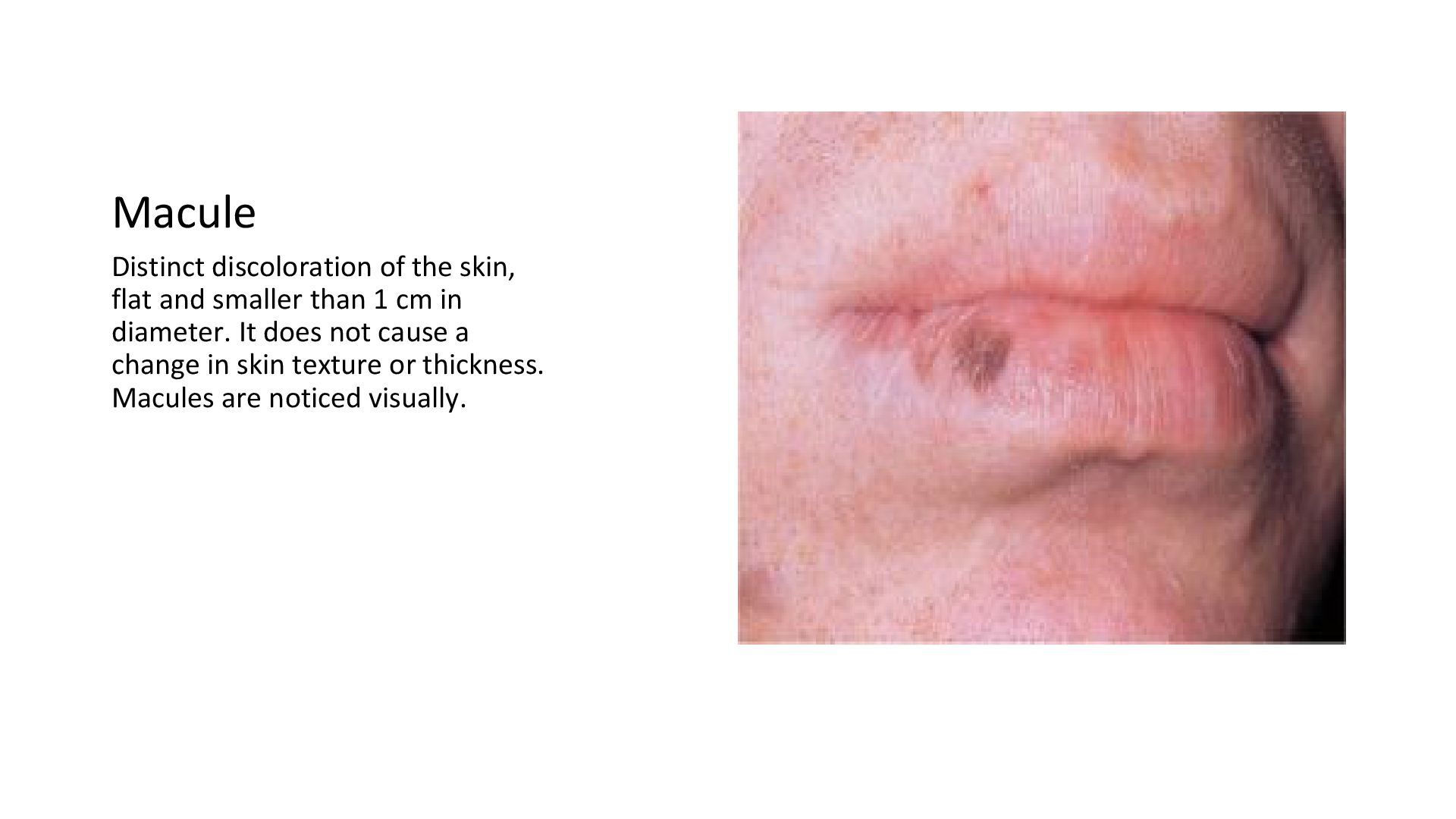
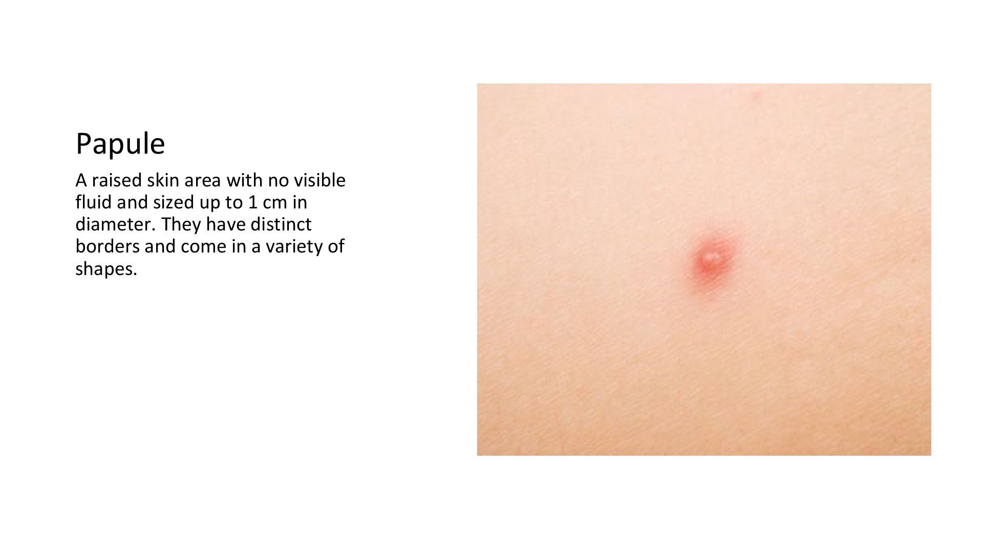
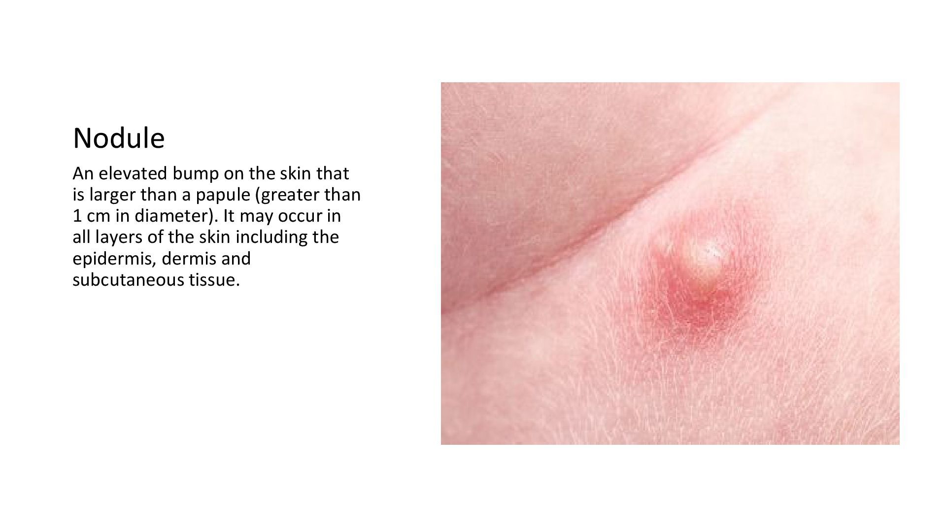
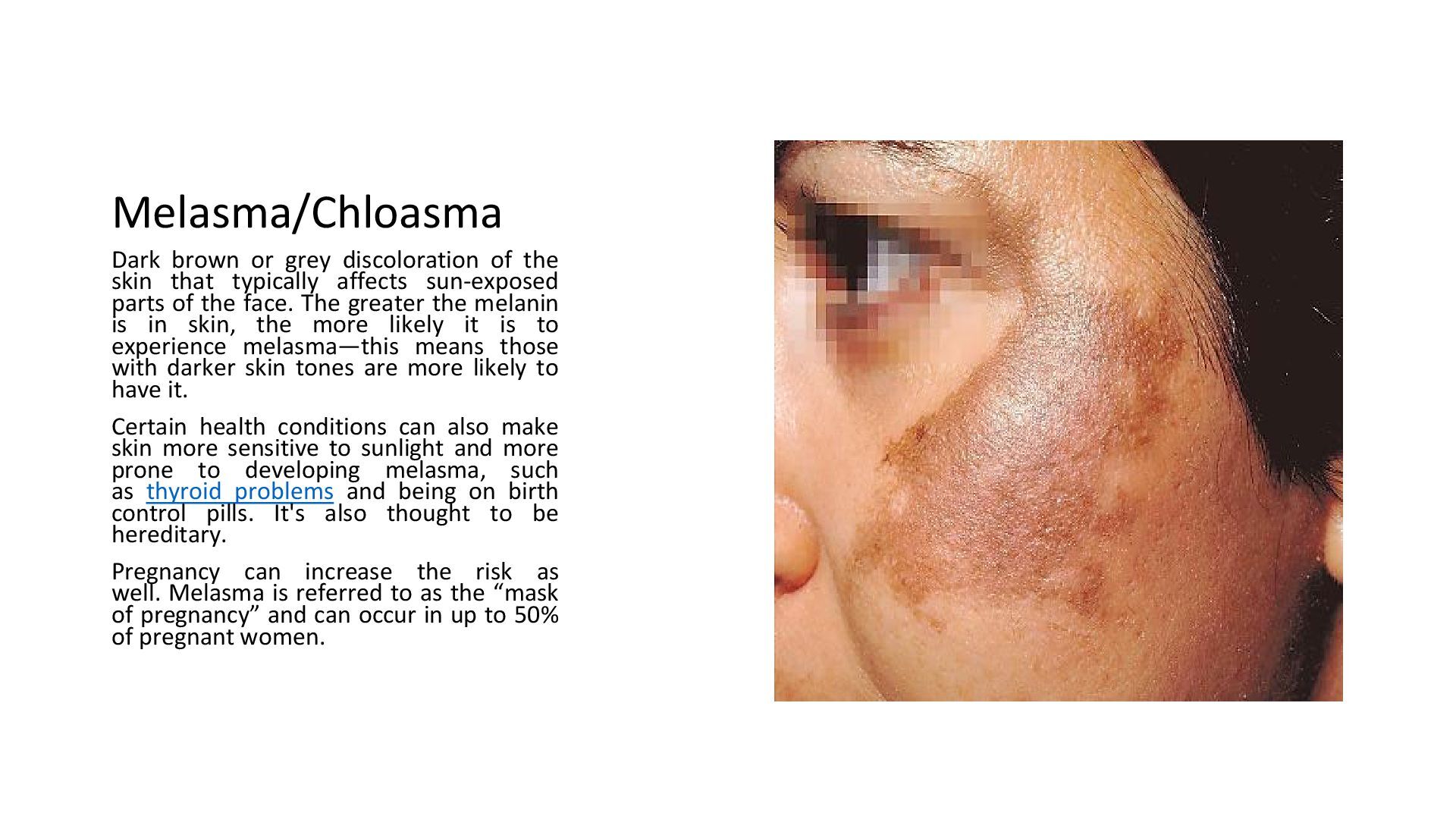
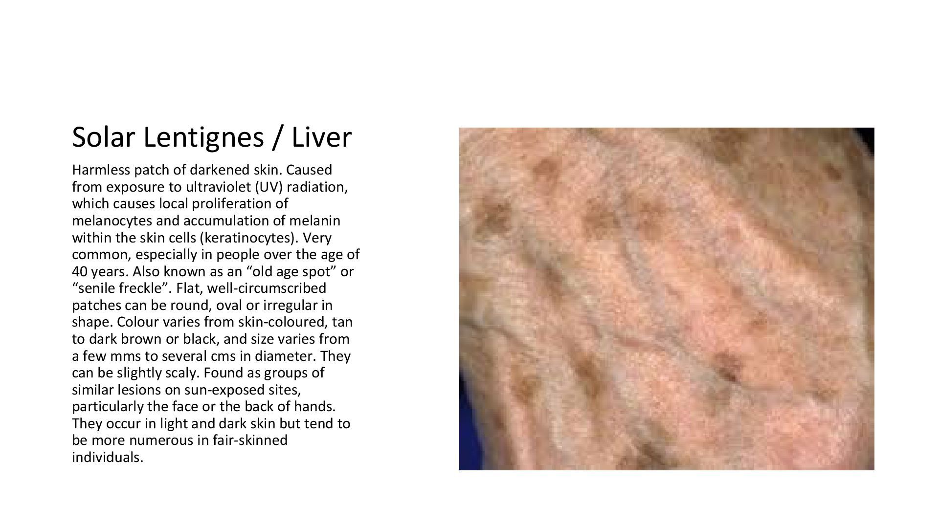
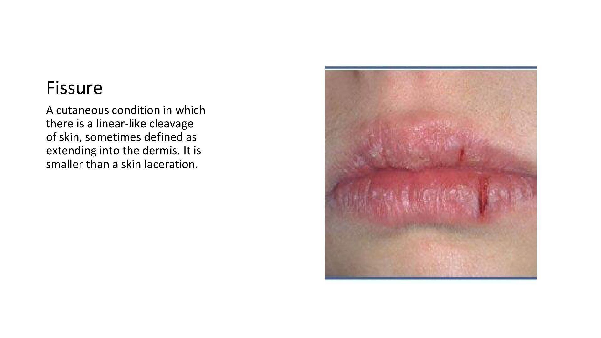
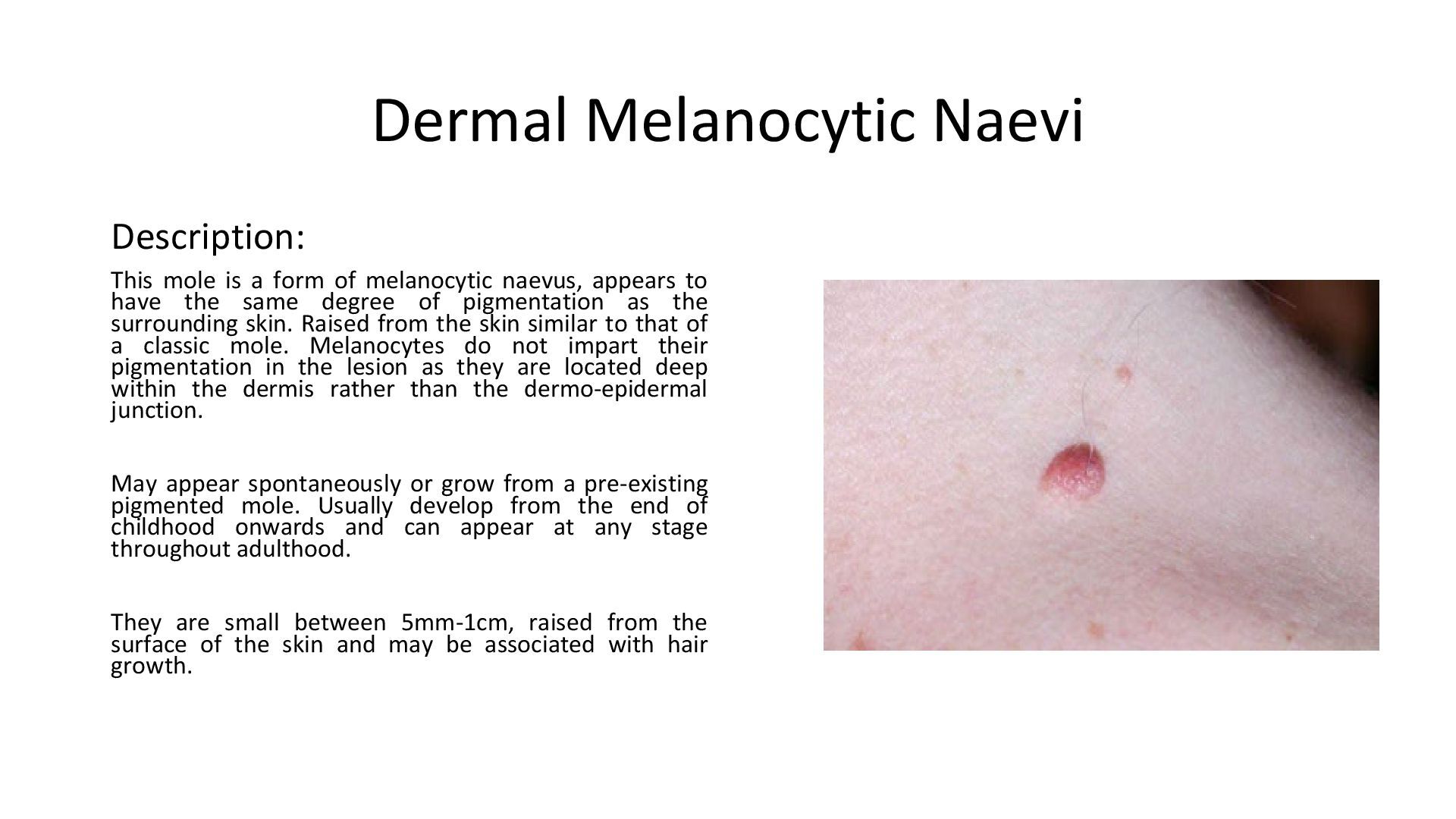
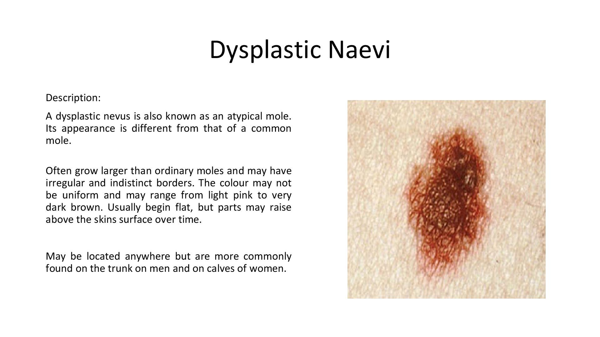
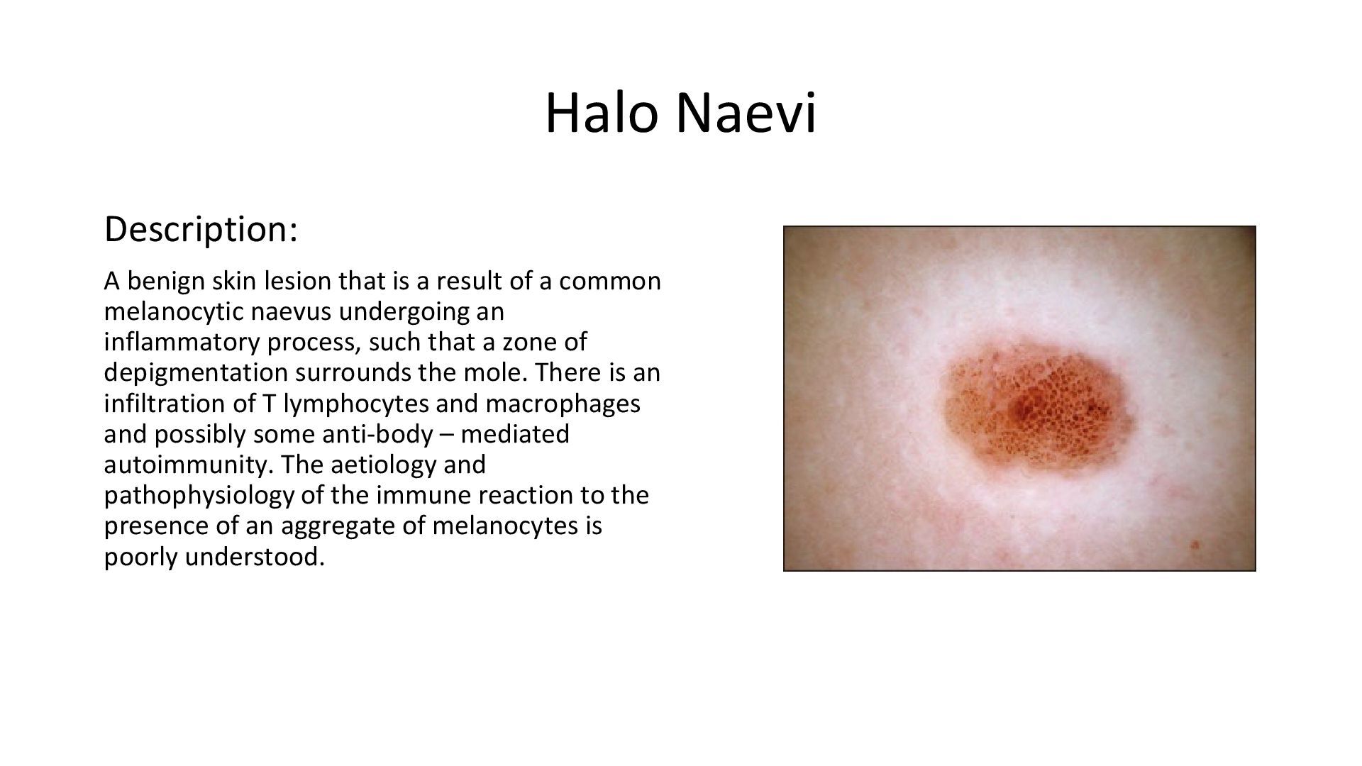
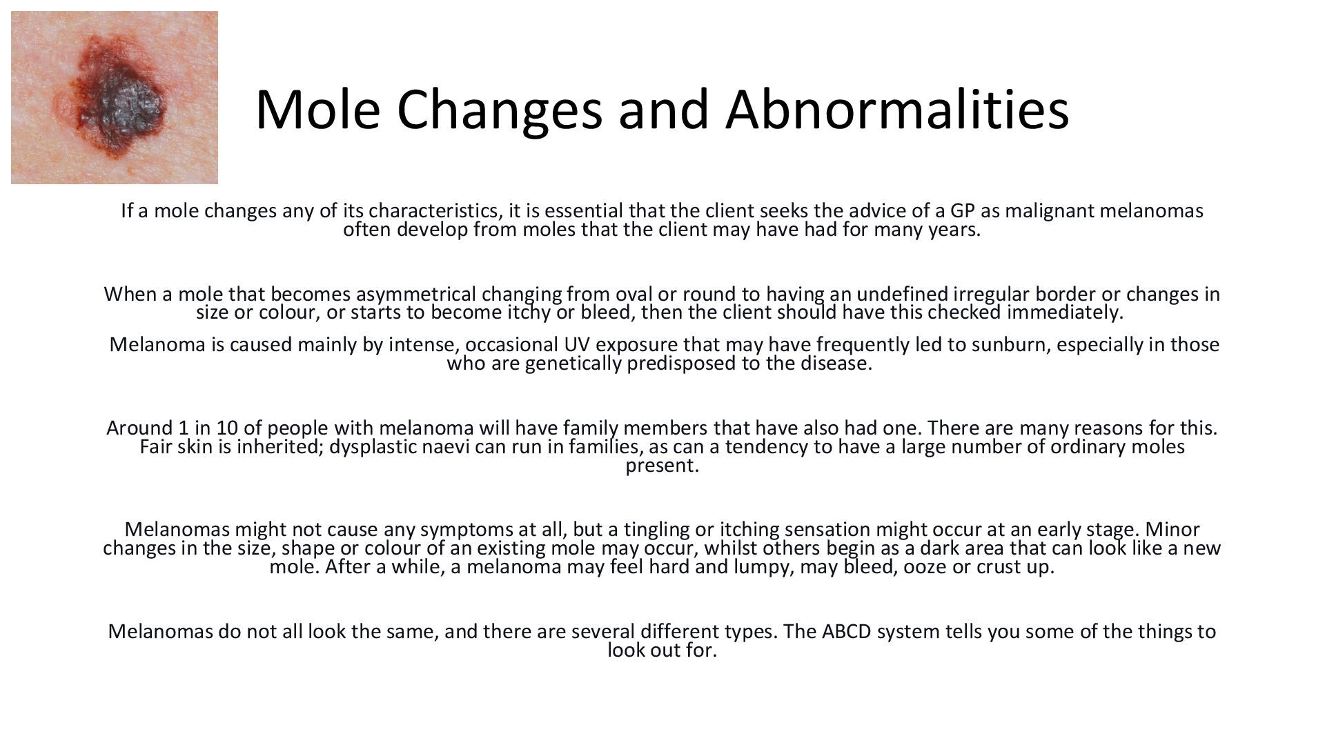
    is Squamous cell carcinoma (SCC). This is usually found in areas of the body that has been damaged by UV rays from the sun or tanning beds. Sun-exposed skin includes the head, neck, ears, lips, arms, legs, and hands. CC is a fairly slow-growing skin cancer. Unlike other variants of skin cancer, SCC can spread to the tissues, bones, and nearby lymph nodes, where it may become harder to treat. If caught early, it’s easier to treat. Certain things make it more likely to develop SCC: ⮚ Old age ⮚ Men have a higher risk of SCC ⮚ Those that are Fair-skinned ⮚ People with Blue, green, or grey eyes ⮚ Those with Blonde or red hair ⮚ Someone that has spent a lot of time ⮚ outside; exposed to the sun’s UV rays ⮚ Anyone that has used tanning beds and bulbs ⮚ Long-term exposure to chemicals such as arsenic in the water ⮚ Anyone with Bowen’s disease, HPV, HIV, or AIDS ⮚ Previous exposure to radiation ⮚ Inherited DNA condition SCC usually begins as a dome-shaped bump or a red, scaly patch of skin. It’s usually rough and crusty and can bleed easily when scraped. Large growths may itch or hurt. It may also pop through scars or chronic skin sores; clients should get it checked out by their GP immediately.
  42. Basel Cell Carcinoma Basal cell carcinoma is a cancer that

    occurs in parts of the body that gets exposed to a lot of sun. It’s the least risky type of skin cancer; providing that it is caught early, it can be cured. Basel cell carcinoma is unlikely to spread from the skin to other parts of the body, but it can affect nearby bone or other tissue under the skin. Several treatments can prevent that from happening and get rid of cancer. The tumour starts off as small shiny bumps, commonly found on the nose or other parts of the face. However, you can get them on any part of your body, including your trunk, legs, and arms. If the client has fair skin, then they are more than likely to get this skin cancer. Basal cell carcinoma usually grows very slowly and doesn’t show up for many years after exposure to intense or long-term exposure to the sun. It could occur in younger years if the client was exposed to a lot of sun or used tanning beds. Ultraviolet (UV) rays from the sun or from a tanning bed are the main causing factor of basal cell carcinoma.
  43. Basel Cell Carcinoma When ultraviolet rays penetrate the skin, over

    time, they can cause damage to the DNA in the skin cells. The DNA holds the code for the way these cells grow. Over time, this damage to the DNA can cause cancer to form. The process can take many years. Basal cell carcinoma can look different. Your clients may notice a skin growth in a dome shape that has blood vessels in it. It can be pink, brown, or black. In the start, a basal cell carcinoma comes up like a small “pearly” bump that looks like a flesh-coloured mole or a pimple that doesn’t go away. Sometimes these growths can look dark. Or the client may also see shiny pink or red patches that are slightly scaly. Another symptom to watch out for is a waxy, hard skin growth. Basal cell carcinomas are also fragile and can bleed easily.
  44. Haemangiomata – Cherry Angiomas – Campbell De Morgan Spots Angioma

    or haemangioma is the name of a benign or non-cancerous overgrowth of blood vessels in the skin. Angiomas in adults include cherry angiomas or more commonly known as blood spots or Campbell de Morgan spots, and they may be found on any part of the body, but more commonly, they appear mid-torso. They can be red, purple, blue or almost black in colour and they generally increase in number from the age of 40. Haemangiomata’s are most likely caused by the proliferation of endothelial cells that line the blood vessels, and whilst the cause is still unknown, it seems to be part of the natural ageing process.
  45. Skin Tags Skin tags are harmless and benign skin growths

    that are commonly found on the neck, axilla region, under the bra area and in the groin. They appear in various colours, from flesh to dark brown. They may be just slightly raised, or they will have a stalk or neck like a mushroom, or they might have a flat neck. They are very common, and they may be a treatment you perform often. Clients that develop several skin tags may have a genetic predisposition as this condition often runs in families. They often start appearing with age, especially in people over 50 years of age. Friction is also another contributing factor as they appear in the areas where clothes may rub, such as collar lines, bra straps, underarms and groin etc.
  46. Common Warts Common warts are small rough, hard, nodular growths

    on the skin and develop due to an over- accumulation of hyper keratinised cells. They can occur singly, although they are more commonly found in groups on the hands, fingers or knees, but they can be found anywhere. Anyone can suffer from common warts, but they are mostly found in children. Warts may present in several forms. Flat or plane warts can appear on the legs or face and often appear in groups. Perifungal warts refer to warts that are found under nails or around them, filiform warts appear on the face like single long stalks, and plantar warts are found on the bottom of the feet, more commonly known as verrucae. Common warts are caused by a viral condition called the Human Papilloma Virus, and whilst they are not harmful, they are contagious. If the virus is active, they will be difficult to treat successfully with any method. A referral to a GP may be required, and therapists should check the parameters of their insurance.
  47. Seborrheic Warts or Keratosis Seborrheic keratosis, seborrheic warts and basal

    cell papilloma’s and are very common, harmless, usually pigmentated, skin growths. They are benign growths caused by a build-up of keratinocyte cells. They start to appear usually from the age of 40 years, although they may appear in younger people, however, the majority of older people will have a few seborrheic keratoses, while some may have quite a lot. Seborrheic keratoses have a rough surface and can range in colour from light to dark brown to almost black. Small flat seborrheic keratosis may become more raised and larger as the years go by. Their size can vary from less than one centimetre to several centimetres across. They can give the impression that they are attached to the surface of the skin; some may look like small pigmented skin tags. Seborrheic keratosis is most commonly found on the trunk, but they can also be seen on the head and neck. Numbers may vary, and once present, they usually stay, and new ones can appear over the years. They can affect anyone, but on darker skin, they can appear as multiple small dark brown or black bumps, especially on the face and neck and are known as dermatosis papulosa nigra. Seborrheic keratosis is often referred to as warts, but they are not caused by the wart virus. Age is a contributory factor as a result of an overproduction of keratinocytes. They are not malignant or contagious and can be treated. A GP referral or approval should be sought as they can often look similar to melanoma. Treatment with electrical currents can be very successful, although darker skin tones should be treated with caution as removal may result in hyperpigmentation.
  48. Solar lentigo Solar lentigo (a type of lentigo also known

    as a senile lentigo, age spots, or liver spot) is a benign pigmented macule appearing on fair-skinned individuals that is related to ultraviolet radiation (UV) exposure typically from the sun. Solar lentigines are believed to be UVR-induced proliferative responses of the epidermal keratinocytes and/or melanocytes, although the exact mechanism of formation is not completely understood. Ultraviolet B (UVB) exposure is thought to increase the expression of keratinocyte growth factor, which thereby induces tyrosinase expression and melanin production in melanocytes. This melanin pigment is then transferred to keratinocytes, where there is abnormal pigment retention. Solar lentigines are more common in those with skin phototypes I-III and a history of multiple sunburns. They are present in 90% of individuals over 60 years of age who are of Northern European descent. They are typically located on sun-exposed skin, including the face, upper chest, shoulders, dorsal arms, and hands. Solar lentigines are asymptomatic, although they may enlarge, darken, or remain unchanged over time.
  49. Loading the Cryopen ⮚ Before loading a new cartridge, release

    any unused gas from the current cartridge by unscrewing the applicator followed by pressing the button. Then unscrew the device. Discard the used cartridge. Ensure that the used filter does not remain inside the cavity. ⮚ The cartridges are packed in peel-pack blisters with an assembled filter and cartridge opener included. The filters are used for the protection of the applicator, the opener for puncturing the cartridge. Ensure that the loading cavity is empty of all residue. Open the peel-pack. Take out the filter/opener with the tweezers. Hold the CryoPen tip end pointed downward. Place the filter in the instrument and make sure that it is seated flat at the bottom of the loading cavity. The trocar of the opener should be pointed upwards. Take the cartridge and put it into the cavity with the narrow side down, on top of the filter/opener device. ⮚ Gently screw the CryoPen’s backend onto the front end. Go to the point where you feel that the contact between the assembled filter/opener device and the cartridge is established. Position your hands so that the last turn will be in an uninterrupted movement. Rotate quickly until you feel that the end of the rotation is achieved. You may eventually hear a short hissing sound. The shorter the hiss, the better. ⮚ Remove the applicator cap. Gently squeeze the trigger switch to start the flow of N2O. Release the trigger to close off the flow of cryogenic liquid. 1 or 2 seconds after releasing the trigger, the remaining gas in the applicator is discarded.
  50. Cryopen Health & Safety ⮚ Always gently manipulate all components

    of the CryoPen instruments. Never use more than gentle hand force. Never use pliers or other tools to manipulate the instrument. ⮚ Ensure to only use the filter/opener supplied with the N2O cartridges. Any other filter/opener would damage the pen. ⮚ Incorrect placement of the filter/opener device into the loading cavity will cause irreversible damage to the instrument. ⮚ Residual foreign matters in the loading cavity while placing a new filter/opener can cause instrument failure. ⮚ When loaded, the instrument is under high pressure. Any change to the construction of the instrument may cause a potential risk. ⮚ Never put your hand on the opening while removing the cartridge. ⮚ All external parts can be wiped with a cloth soaked in any noncorrosive disinfecting solution. Never immerse the entire instrument in a disinfecting solution. Only the applicators can be autoclaved. ⮚ The applicators and the cartridges must be kept away from dust and high temperatures. Leave the cap on when not in use. ⮚ The CryoPen should be handled with care. When storing the instrument with a loaded cartridge in its case, position the CryoPen in such a way that the trigger switch cannot be depressed when closing the lid of the case. This would cause undesired gas flow. ⮚ The N2 O cartridges contain both a liquid and a gaseous phase in balance. The proportion of each phase depends on the room temperature, which affects the output flow. The best performances are achieved when the cartridge temperature is between 19°C and 22°C (66°F and 72°F). Lower and higher temperatures will give different values in respect of pressure and the density of the cryogen. Over 25°C/77°F, the quantity of liquefied N2 O in the CryoPen jet will decrease. ⮚ Empty cartridges should be discarded as metal scrap.
  51. Treatment Considerations Before treatment begins, a number of factors should

    be taken into consideration. Ensure the client is suitable for treatment by undertaking a full client consultation to ascertain if there are any contraindications present and what type of blemish or lesion is to be treated. You may need to seek GP permission for certain blemishes or where there is uncertainty on the type of blemish or lesion that needs to be treated. Check with your insurers to ensure that you are covered for all treatments you wish to perform.
  52. Contra-Indications A contraindication is the presence of a condition which

    may make the client unsuitable for the treatment. The treatment may not be able to take place, or the treatment will need to be adapted. When treating a client, if they show any signs of contra-indications, you should tactfully refer them to their GP for treatment or advice. You should never make a diagnosis even if you are certain of the condition as you may be wrong. If you are unsure about any contra-indications, then do not treat the client and refer them to their GP. Be careful if you deal with a contra-indication, and they can often be contagious. Make sure you clean the work area and any implements between clients to prevent cross-infection.
  53. Diabetes: Description: Diabetes is a lifelong condition that causes a

    person's blood sugar level to become too high. There are two main types of diabetes: type 1 diabetes – where the body's immune system attacks and destroys the cells that produce insulin type 2 diabetes – where the body does not produce enough insulin or the body's cells do not react to insulin. Type 2 diabetes is far more common than type 1. In the UK, around 90% of all adults with diabetes have type 2. The amount of sugar in the blood is controlled by a hormone called insulin, which is produced by the pancreas (a gland behind the stomach). When food is digested and enters the bloodstream, insulin moves glucose out of the blood and into cells, where it's broken down to produce energy. However, with diabetes, the body is unable to break down glucose into energy. This is because there's either not enough insulin to move the glucose, or the insulin produced does not work properly. Salon Treatment: Can often be treated if the client is managing their diabetes well. Check with your insurer as their criteria may not allow treatment. A GP note may be required. Treatments that cause injury to the skin can increase the risk of infection. Those with diabetes will need to understand wound management and treatments should be done under extreme caution as to not injure the skin if necessary. Diabetics have a slower wound healing response and have a higher risk of infection. All products used must be sterile, and care should be taken not to injure the skin.
  54. Epilepsy: Description: Epilepsy is diagnosed when a person has had

    more than one epileptic seizure and could have more in the future. Electrical activity is happening in our brain all the time. A seizure happens when there is a sudden burst of intense electrical activity. This is often referred to as epileptic activity. This intense electrical activity causes a temporary disruption to the way the brain normally works, meaning that the brain’s messages become mixed up. The result is an epileptic seizure. There are many different types of seizure, and each person will experience epilepsy in a way that is unique to them. Some things make seizures more likely for some people with epilepsy. These are often referred to as ‘triggers. Triggers are things like stress, not sleeping well and drinking too much alcohol. Some people say they have more seizures if they miss meals. Not taking epilepsy medicine is another common trigger. A very small number of people with epilepsy have seizures triggered by lights that flash or flicker. Avoiding triggers can stop them from having seizures. Salon Treatment: If well-managed treatments may be able to be undertaken. It is worth assessing the client and finding out what triggers a fit and when as well as how it is managed and their last episode. You may also need to check your insurer's terms and refer the client to a GP for a letter of approval.
  55. Heart Conditions: Description: Heart disease describes a range of conditions

    that affect the heart. Heart diseases include: ⮚ Blood vessel disease, such as coronary artery disease ⮚ Heart rhythm problems (arrhythmias) ⮚ Heart defects you're born with (congenital heart defects) ⮚ Heart valve disease ⮚ Disease of the heart muscle ⮚ Heart infection Risk of fainting, risk of slow healing or chances that the client may be medicated to keep their condition under control are some risks you will need to be aware of when treating the client. Clients may also have a heart catheter or a pacemaker. Salon Treatment: Treatments that do not involve electrical currents being passed through the body are usually safe. Heart conditions should be well managed. You will need to check the terms of your insurance policy. A referral to a GP may be required.
  56. Deficient Immune System: Description: Immunodeficiency disorders prevent the body from

    fighting infections and diseases. This type of disorder makes it easier to catch viruses and bacterial infections. Immunodeficiency disorders are either congenital or acquired. A congenital, or primary disorder is one you were born with. Acquired, or secondary, disorders you get later in life. Acquired disorders are more common than congenital disorders. Your immune system includes the following organs: ⮚ spleen ⮚ tonsils ⮚ bone marrow ⮚ lymph nodes These organs make and release lymphocytes. These are white blood cells that fight invaders cells called antigens. Cells release antibodies specific to the disease the body detects. White blood cells destroy foreign or abnormal cells. Examples of antigens that white cells might need to fight off include: ⮚ bacteria ⮚ viruses ⮚ cancer cells ⮚ parasites An immunodeficiency disorder disrupts your body’s ability to defend itself against these antigens. Salon Treatment: A deficient immune system can increase the risk of infection. Treatments that break the skin may increase the risk of infection. Clients may need a GP referral and understand wound management.
  57. Pregnancy: Description: Pregnancy is a period of considerable changes in

    a woman's body. These changes, affecting virtually every part of the body, are all geared towards growing and delivering a healthy baby, without harming the mother. Changes begin within days of conception when the fertilised egg implants itself in the wall of the uterus. The first changes are subtle, and most women will not notice them. Pregnancy lasts an average of 266 days (38 weeks) from the date of conception or 40 weeks from the first day of the last menstrual period. Pregnancy is divided into three periods of three months each. These periods are known as the first, second and third trimesters. Each trimester brings with its own unique set of experiences. Risks of pregnancy include: ⮚ Miscarriage ⮚ Increased/decreased blood flow ⮚ Risk of fainting ⮚ Oedema ⮚ Pigmentation/Chloasma/mask of pregnancy ⮚ Pelvic trauma Salon Treatment: Nearly all treatments are contraindicated in the first trimester. Treatments that cause friction may have a side effect of PIH and should not be performed. Electrical treatments must not be performed at any point.
  58. Breastfeeding: Description: Anything the mother consumes or applies to her

    skin could be absorbed into the bloodstream and through into the milk ducts, where this can pass to the baby. Great care should be taken with what is being applied to the skin as well as avoiding treatments that could cause open wounds. An open wound is susceptible to infection, and this can cause infection or septicaemia. During breastfeeding, hormones are still active within the body, and these can have an overall effect on the skin. This may lead to dehydrated skin (if the mother is not ingesting enough water) and also lead to a high risk of pigmentation. Invasive procedures should be avoided until a few months after the mother has stopped producing milk. Salon Treatment: Treatments that break the skin are strictly contraindicated. The client also has a high amount of hormone imbalance still and may be susceptible to PIH on certain treatments.
  59. Electrical implants or pacemakers: Description: A small battery-operated device called

    a pacemaker is placed into the chest. It sends regular electrical impulses, which help keep the heart beating regularly. The pacemaker is a small metal box weighing 20–50g. It is attached to one or more wires, known as pacing leads, that run to the heart. If the pacemaker senses that the heart has missed a beat or is beating too slowly, it sends signals at a steady rate. If it senses that the heart is beating normally by itself, it does not send out any signals. Most pacemakers have a special sensor that recognises body movement or the breathing rate. This allows them to speed up the discharge rate when active. An implantable cardioverter-defibrillator (ICD) is a device similar to a pacemaker. An ICD delivers an electrical shock to the heart during a life- threatening heart rhythm. The aim is to 'reboot' the heart to get it back into a normal rhythm again. Some modern devices contain both a pacemaker and an ICD. Salon Treatment: Electrical implants contra-indicate any treatment that involves an electrical current. Other treatments that may affect blood pressure may also contra-indicate a procedure. Check your insurer's terms and refer the client to GP for a letter of approval.
  60. Anticoagulant medicines such as warfarin or aspirin: Description: Anticoagulants are

    medicines that help prevent blood clots. They're given to people at a high risk of getting clots, to reduce their chances of developing serious conditions such as strokes and heart attacks. A blood clot is a seal created by the blood to stop bleeding from wounds. While they're useful in stopping bleeding, they can block blood vessels and stop blood flowing to organs such as the brain, heart or lungs if they form in the wrong place. Anticoagulants work by interrupting the process involved in the formation of blood clots. They're sometimes called "blood- thinning" medicines, although they don't actually make the blood thinner. Although they're used for similar purposes, anticoagulants are different from antiplatelet medicines, such as low-dose aspirin and clopidogrel. Salon Treatment: Anticoagulants prevent the blood from clotting and can affect the healing process of the skin. The client will be at higher risk of bruising also. Treatments that injure the skin or cause trauma will need to be avoided. A GP letter may be required for some services.
  61. Steroids, antidepressants, antibiotics and other medications: Description: Many drugs can

    affect the skin. For example, prescription antidepressants, especially the popular class of medications called selective serotonin reuptake inhibitors (SSRIs), can cause a wide variety of adverse skin reactions, including bruising. Acne medications and certain antibiotics may make the skin more sensitive to sunlight and light or heat-based procedures. It is vital that we find out the medications both prescription and over the counter drugs that the client is taking. The internet is a great source of information to establish the risks the medication may have on the skin during a procedure. Salon Treatment: Medications can have numerous effects on the skin. If you are unsure, you should ascertain how the treatment affects the skin and the effects of the medication that is being taken. A patch test may be required or a letter from the clients GP. Where the side effects may be photosensitivity or thinning of the skin, then treatments should be avoided until the client has come off their medication for three months at their GPs request.
  62. Recent Surgery: Description: Clients will have surgery for a variety

    of reasons and on any area of the body. Surgery puts the body under extreme pressure. The client may be on pain medication which could affect their susceptibility to feeling pain during treatment. Surgery wounds can take many months to fully heal. Post-surgery complications can also arise a few months after the procedure. This can include, infection or necrosis of the wound, infection of the blood, blood clots, internal bleeding, or fever. Salon Treatment: If the client has had recent surgery, you will need to weigh up the risk of the procedure, the length of time between the surgery and treatment as well as where the surgery was. In most cases, it is advised to wait six months post-surgery before undertaking any procedures, especially where these may involve invasive techniques, massage or electrical machines.
  63. Metal Plates or Pins: Description: If you break a bone,

    it will need to be held in place while it heals. This can be done using a splint, sling, brace or cast. But sometimes a broken bone will need to be fixed with surgery, using metal plates, rods, wires, screws or pins. Implanted metal can help broken bones heal in proper alignment. While these implants do not help the bone heal faster, they can help to hold bones in the proper position while healing takes place. Implants may include metal plates and screws, pins, and intramedullary rods inserted into the cavity of a bone. Salon Treatment: Electrical treatments are almost always contraindicated for clients with metal implants in their body. However, this can vary depending on the area of the implant vs the area of treatment. Care may also need to be taken when massaging over the area or any pressure is applied.
  64. Chemotherapy/ radiotherapy: Description: Chemotherapy is a form of cancer treatment

    where a patient is given drugs designed to kill cancer cells. Radiation is a type of cancer treatment where high doses of radiation are delivered to cancerous tumours in the body. Both procedures can affect the sensitivity of the skin, and the skin texture may change. A waxy appearance to the skin can alter the effects of treatments and treatments may not be suitable. Clients may also be at risk of pigmentation. Salon Treatment: Clients undergoing treatment for cancer may benefit from a number of procedures in a salon for the purpose of relaxation and pain management as well as to make them feel good. Some treatments may be too much for the client to handle and cause unwanted side-effects. It is worth discussing the treatment with the client and working out alternatives where suitable. If unsure, you may wish to ask for a letter from the client's oncologist.
  65. Broken Bones: Description: When a bone has an outside force

    exerted upon it, like a blow or a fall, there is potential that it cannot withstand the amount of force and it breaks. That loss of integrity results in a fracture. It is important to remember that a fracture, break, or crack all describe the same situation, an injury to the bone where it has been damaged. One term is not more serious than another. Fracture break and crack all mean the same thing. Salon Treatment: Clients with a suspected or recent fracture should be treated with caution. If working over the area of a recent break, then the time between the break and the treatment should be at least six months. If working on other areas of the body, it is best to wait for 6-8 weeks post- injury as the client may be more susceptible to an embolism.
  66. Previous or recent treatments in the area: Description: When providing

    a service to the client, it is important to establish what previous treatments they have had, not only at clinics but at home. You should look to enquire over the products they use regularly that may affect the skin and the treatment outcome. Salon Treatment: Some treatments can affect the skin by either removing out layers of the stratum corneum or stimulating a healing response. You will need to identify if the client has had any recent procedures in the treatment area you will be working on.
  67. Psoriasis • Description and Cause: Scaling and inflammation of the

    skin. Cause unknown but thought to be related to the nervous system • Salon Treatment: Treat with caution; avoid the affected area. Do not treat if the skin is weeping.
  68. Eczema • Description and Cause: Atopic eczema is a common

    skin condition that causes patches of skin that are itchy, cracked and sore. • Salon Treatment: Treat with caution: avoid the affected area.
  69. Conjunctivitis • Description and Cause: A transparent and sticky substance

    covers the white of the eye and lids. It is caused by a bacterial infection. • Salon Treatment: This is highly infections; do not treat; the client should be referred to their GP for correct diagnosis and treatment.
  70. Cuts & Abrasion • Description and Causes: Broken skin caused

    by an injury. • Salon Treatment: Avoid treatment in the affected area.
  71. Bacterial Keratitis • Description and Causes: A severe disorder which

    can result in partial or total loss of vision. It is caused by a bacterial infection. • Salon Treatment: Do not treat; refer clients to their GP for correct diagnosis and treatment.
  72. Bruising • Description and Causes: Black, green. Yellow or red

    marks appear on the skin. They are generally caused by an injury. • Salon Treatment: Avoid the area if possible.
  73. Recent Operations (Scars) • Descriptions and Causes: Scar tissue raised

    or flat undergoing the healing process. Scar tissue is very sensitive. • Salon Treatment: Avoid treatment if the scar is less than six months old.
  74. Blepharitis • Description and Causes: Inflammation of the eyelids; the

    inflammation is like eczema of the skin with red, scaly eyelids; you may notice tired or gritty eyes, which may be uncomfortable in sunlight or smoky atmospheres; they may be red and swollen and feel as though there is something in them. The exact cause is unknown, but people who have dandruff or dry skin conditions may be more prone to blepharitis. • Salon Treatment: Avoid the area; refer the client to GP for correct diagnosis and treatment.
  75. Stye • Description and Causes: Infection in the root of

    an eyelash. They are caused by a bacterial infection. • Salon Treatment: Avoid the area; no treatment until the infection has gone.
  76. Sunburn • Description and Causes: Sunburn is a red, painful

    skin that feels hot to the touch. It usually appears within a few hours after too much exposure to ultraviolet (UV) light from sunshine or artificial sources, such as sunbeds. • Salon Treatment: No treatment should be provided until the skin has healed. 48 hours should be left from sun exposure/sunbed use before a treatment.
  77. Cold Sores • Description and Causes: Cold sores are painful

    lumps or blisters on the face. They're caused by a virus and are very contagious. • Salon Treatment: Treatment should not be provided on the face until the skin has healed. Precautions should be taken when treating other areas to avoid cross-contamination.
  78. Impetigo • Description and Causes: Impetigo is a highly contagious

    skin infection that often starts with red sores on the face or hands. It causes red sores or blisters that burst and leave crusty, golden-brown patches. • Salon Treatment: Treatment should not be provided on the face until the skin has healed. Precautions should be taken when treating other areas to avoid cross-contamination.
  79. Moles • Description and Causes: Moles are small, coloured spots

    on the skin. Most people have them, and they're usually nothing to worry about unless they change size, shape or colour. • Salon Treatment: Avoid treating over a mole. Irritation or damage to the area may cause the mole to change.
  80. Ringworm • Description and Causes: Ringworm is a fungal infection

    on the skin. It causes a rash that is often ring-shaped. • Salon Treatment: Treatment should not be provided to the client until the condition has been treated.
  81. Sensitive Skin • Description and Causes: Sensitive skin is a

    common issue but not a medical diagnosis in itself. The term generally refers to skin that is more prone to inflammation or adverse reactions. People with sensitive skin may have strong reactions to chemicals, dyes, and fragrances present in products that come into contact with the skin. • Salon Treatment: Treatments should be applied with caution. Patch tests may be required before undertaking a full procedure.
  82. Other Contra-indications to Treatment ⮚Unstable diabetes ⮚Skin conditions, e.g. skin

    tumours, open wounds, solar hyperkeratosis ⮚Unexplained, suspicious liver spots and moles ⮚Cancer tissues and malignant tumours
  83. The contraindications related to temporary conditions: ⮚Infections accompanied by fever

    ⮚Acute chemotherapy or radiotherapy from four weeks before beginning the therapy to four weeks after finishing the therapy ⮚Pregnancy or breastfeeding ⮚Cold intolerance ⮚Vascular insufficiency
  84. Although cryotherapy is a relatively low-risk procedure, some side effects

    may occur as a result of the treatment. They include: ⮚ Permanent changes in pigmentation. Both hypopigmentation (lightening of the skin) and hyperpigmentation (darkening of the skin) may occur after cryotherapy. Both generally last a few months but can be longer- lasting. Avoid freezing the basal cell layer where melanocytes (pigment-producing cells) are located. ⮚ Sensory impairment. Though rare, damage to nerves is possible, particularly in areas where they lie closer to the surface of the skin, such as the fingers, the wrist, and the area behind the ear. Reports suggest this will disappear within several months. ⮚ A spattering of the cryogen during spraying, when the end nozzle freezes. The innovation of CryoPen is the direct application of nitrous oxide under high pressure (55 bar). This high-pressure jet may cause minor shards of frozen humidity in the air blown away in a circle of approximately 30cm in diameter. They will thaw the moment they would eventually touch healthy skin. ⮚ Hair loss. Hair follicles are easily damaged by cryosurgery, and permanent hair loss is not uncommon. ⮚ Pigmented spots. It is common use to not treat pigmented spot in sunny seasons. Solar lentigines (SL) are flat brown sharply demarcated pigmented lesions that commonly appear as a sign of ageing and photodamage on sun-exposed skin. ⮚ Post-inflammatory hyperpigmentation (PIH) is a frequent concern when treating SL in darker skin types.
  85. Further Side Effects: ⮚ Edemas ⮚ Bleeding ⮚ Pain and

    syncope ⮚ Insufflation of subcutaneous tissue ⮚ Retraction caused by freezing next to orifices ⮚ Cartilage defect during treatment of the nose or the ear ⮚ Infection ⮚ Conjunctival ectopy due to treatments of the eyelid ⮚ Milia ⮚ Depressed or atrophic scars
  86. Client Consultation A client consultation is a one to one

    talk with your client. During this time, you will find out very important and confidential information that will allow you to advise and provide the best treatment for the client. It is important to always introduce yourself to the client as this removes any barriers and relaxes them. Consultations should always be undertaken in a private room or area where you cannot be overheard by others. A client should first fill out a client consultation which helps identify any contra-indications that may mean you have to alter the treatment or be unable to treat them at all. If their form shows no reason why they cannot proceed with the treatment, then you can move onto verbal questioning. Verbal questions would be to establish why the client has visited the salon and what their expectations and outcome of the treatment may be. Asking what they want ensures you can provide customer satisfaction as the client should be pleased with the outcome of their treatment. It is good practice to speak to the client in front of a mirror and explain the treatment to them and see if that meets their requirements.
  87. Client Consultation Once you have established what the client is

    after, then a physical examination should be undertaken. This allows you to further check for any undeclared contra-indications and get a better overview of any issues that you may face during the procedure. Allow around 15 minutes for the client's first salon visit. Ideally, you should sit face to face or next to the client to create an open atmosphere. Avoid barriers such as a couch or table between you. Use open questions to tactfully encourage the client to give you information that you may need rather than using interrogating questioning techniques. Use the consultation form to work from and record anything you may discuss.
  88. Record Keeping Records must be maintained and updated for a

    number of reasons. ⮚ They provide contact details in case you need to alter or cancel an upcoming appointment. ⮚ So that you can track client’s progression. ⮚ To record the products used and timings so you can use these at further visits and adjust the treatment plan if required. ⮚ Tracks any aftercare you provide the client. ⮚ Records patch test history. ⮚ As a backup in case, the client has an adverse reaction to treatment. ⮚ For legal reasons if the client brings a claim against you.
  89. Record Keeping Client records can be stored electronically or filed

    away manually and should be updated at every visit. If consultation forms are not updated and do not contain a history of services and dates, then you may find your insurance invalid. Forms should be kept for the timeframe suggested by your insurance company. This may be for up to six years. If a client is under 21 at the time of service, then it is recommended to keep the forms for six years past their 21st birthday. Client confidentiality must be protected at all times. Forms need to be locked away in a secure cabinet, and electronic records should be held on a password-protected computer. You may also need to register with the ICO as a data controller. All information must be accurate and necessary for the service or treatment being performed. Individual client records must be available for the clients to view if requested. Data should not be passed on or sold without the client’s prior written permission.
  90. Consultation Forms The following details should be recorded on the

    client consultation form: ⮚ Personal details – name, address, contact details ⮚ Results of any patch tests ⮚ Contra-indications ⮚ Contra-actions ⮚ Reasons for the treatment ⮚ Any reactions to treatments/previous treatments ⮚ Home care advice/suggested retail items. ⮚ Any sales ⮚ Treatment timings/products used etc. ⮚ Next appointment or recommendations Any contra-indications and possible contra-actions should be identified and discussed prior to the treatment. In the case of a medical referral, the therapist should keep a copy of the GP's letter with the client's record card. Consultation forms must be signed and dated to prove that you have covered everything and given the correct advice and treatment plan.
  91. Treatment Protocol CryoPen: Treatment Guidelines Before treating, always consider: •

    Client’s age and skin type • Lesion size, type, and number • Location of the lesion • Skin tanning level and the current season
  92. Application Technique: • Hold the applicator 0.5–3 mm from the

    lesion • Do NOT go beyond 5 mm, or the gas becomes ineffective • Move the pen in small, circular or cross-shaped motions • The closer you are, the stronger the freezing effect • For smaller or delicate lesions, maintain more distance • Adjust freeze strength by moving closer or further away during application
  93. Treatment Protocol CryoPen: Application Safety & Positioning Do not hold

    the CryoPen still over the lesion • This may cause excessive freezing, damaging healthy surrounding tissue • Blistering and client discomfort can occur Watch for ice build-up • Air humidity may cause an “ice igloo” to form over the lesion • This layer blocks the effective action of the cryogen • Gently remove with the applicator tip before continuing Aftercare Tip • After thawing, tissue may appear red and inflamed due to minor bleeding • Apply SPF to protect healing skin for 8–20 days after scabbing Positioning Matters • Always hold the CryoPen vertically with the tip pointing down • Do not angle it less than 45°, to ensure precise and safe delivery
  94. Treatment Protocol CryoPen: Treatment Technique & Safety Before You Begin:

    • Consider the age, skin type, tanning level, and number, size, and location of lesions. Application Distance: • Hold the tip 0.5–3 mm from the lesion • Do not exceed 5 mm, or the cryogen becomes ineffective • The closer the tip, the stronger the freezing effect Application Movements: • Move the pen in quick circular or cross motions • For delicate areas, keep more distance and use lighter movements Avoid Over-Freezing: • Don’t hold the pen still – it may flood the area, damaging healthy skin and causing blisters or pain Watch for Ice Build-up: • An “ice igloo” may form from humidity — gently remove it with the tip to maintain freezing power Aftercare Tip: • Treated skin turns red due to inflammation and bleeding • Apply SPF daily for 8–20 days after scab removal to protect healing tissue Pro Tip: • CryoPen offers precise control — practise on common lesions before moving to sensitive areas
  95. CryoPen: Freeze Timing & Technique Application Tips • Hold the

    applicator tip as close as possible to the lesion without direct contact. • Thermal shock (rapid freezing) is essential for success. • Be cautious on areas with thin or sensitive skin. Freeze Duration (Guide Only): • Small, flat wart: 5–10 seconds • Thick plantar wart: Up to 45 seconds • Most lesions: 2–30 seconds depending on thickness
  96. Freeze–Thaw–Freeze Method: 1. Freeze the lesion 2. Allow to thaw

    for 30 seconds 3. Apply a second freeze Second freeze will act faster and is often more effective Clinical Considerations: • Adjust duration based on tissue thickness and pain tolerance • Ice should spread to the base of the lesion before stopping • Mild pain may occur once ice reaches nerve endings • You can extend by a few seconds if the client remains comfortable
  97. Cryotherapy Aftercare Instructions General Advice • Itching for 10 minutes

    after treatment is normal – it’s just histamine response. • Do not pick or scratch the treated area – this delays healing. • If a scab or crust is knocked off, healing may take longer, but results should still be successful. • Blisters may occur (rare with CryoPen) – it’s a known side effect and covered in your consent form
  98. If a blister appears: • Don’t pop it – let

    it heal naturally. • If it bursts: • Gently clean with a non-alcohol baby wipe • Apply Savlon or Germolene twice a day • Use a plaster to protect it if needed
  99. Specific Lesion Aftercare Skin Tags • Will turn black at

    the base and drop off in 1–6 weeks • Larger tags (3mm+) may need a second freeze – contact us after 6 weeks • If sore, apply Savlon or Germolene and cover with a plaster Warts/Verrucae • May require re-treatment after 4 weeks • No aftercare unless a blister forms – follow blister advice Milia • Usually flatten and disappear without any further care • If a blister appears, follow the general blister aftercare Pigmentation/Cherry Angiomas • Area may look raised or like a bee sting – this is normal and will settle in hours • The area will darken and form a crust – do not pick it • When the crust falls off, pink new skin will appear – this is not scarring • Skin will re-pigment naturally over several weeks
  100. Additional Tips • Apply SPF 50 on pigmentation areas until

    normal colour returns (may take up to 8 weeks) • You can use makeup, perfume, or deodorant after treatment – but wait a few hours if possible • If a crust hasn’t fallen off after 4 weeks, be patient – regeneration times vary • Treated tanned skin will lose its tan and return to natural colour • If a lesion hasn’t healed after 6 weeks, consult your GP or dermatologist
  101. Condition Typical Treatment Interval Re-Treatment Notes Warts (common/plantar) Every 4–6

    weeks May require 2–4 sessions depending on size/depth Verrucae Every 4–6 weeks Resistant verrucae may take longer; check progress at each visit Skin Tags Often resolved after 1 session Larger tags (3mm+) may need 1 re- treatment after 6 weeks Solar Lentigines (Age Spots) Usually 1–2 sessions Follow-up in 4 weeks if pigment remains Cherry Angiomas Typically 1–2 sessions Review after 4 weeks Seborrhoeic Keratoses Often cleared in 1 treatment Heavier or thicker lesions may require another session after 4–6 weeks Milia Usually 1 session Reassess after 4 weeks if not fully flattened Actinic Keratoses Every 4 weeks until resolution Monitor closely; GP referral if not resolved after 2–3 treatments Molluscum Contagiosum Every 2–4 weeks May require multiple sessions Cryotherapy Treatment Frequency Guide
  102. General Rule of Thumb: • Allow full healing (crust falling

    off + skin regeneration) before repeating treatment • Do not treat the same site more than once every 3–6 weeks, depending on tissue recovery • Adjust frequency based on client tolerance, lesion size, and location
  103. Condition Freeze Time Notes Common Warts 10–30 seconds Use longer

    times for thicker or older lesions Plantar Warts (Verrucae) 30–45 seconds Often deeper; may require a second cycle after thaw Flat Warts 5–10 seconds Shallow lesions; shorter time is sufficient Skin Tags 5–10 seconds Usually superficial; base will darken and drop off in 1–2 weeks Solar Lentigines (Age Spots) 5–10 seconds Very superficial pigmentation Cherry Angiomas 10–15 seconds Small vascular lesions freeze quickly Seborrhoeic Keratoses 10–20 seconds Time depends on size and thickness Milia 3–5 seconds Very superficial; avoid over-freezing Actinic Keratoses 15–30 seconds Depends on severity and lesion depth Molluscum Contagiosum 5–10 seconds Avoid over-freezing; monitor response Pigmentation (benign) 5–10 seconds E.g. freckles, sun spots Cryotherapy Freeze Duration by Skin Condition
  104. Conditions That Benefit from the Freeze–Thaw–Freeze Cycle Condition Freeze–Thaw–Freeze Recommended?

    Why? Common Warts Yes Enhances effectiveness, especially in older, thicker lesions Plantar Warts (Verrucae) Yes Deep, stubborn lesions require double freeze to penetrate fully Seborrhoeic Keratoses Often Thickened lesions benefit from enhanced cell destruction Actinic Keratoses Yes Precancerous, needs thorough treatment Molluscum Contagiosum Sometimes Use with caution depending on depth; often 1 cycle is sufficient
  105. Conditions That Typically Do Not Require Freeze–Thaw–Freeze: Condition Why Not?

    Skin Tags Superficial; a single short freeze is usually sufficient Milia Very superficial; risk of overtreatment if double freezing used Cherry Angiomas Vascular and shallow; one freeze usually clears the lesion Solar Lentigines / Pigmentation Surface-level melanin – no benefit from a second cycle
  106. Notes: • Always allow 30 seconds of thaw time before

    re-freezing. • Adjust based on lesion size, depth, and client comfort. • Monitor tissue response during first freeze to decide if a second cycle is appropriate.