Upgrade to Pro — share decks privately, control downloads, hide ads and more …

Cryopen-A&P

 Cryopen-A&P

More Decks by Beautiful World Training Academy

Other Decks in Education

Transcript

  1. Skin Anatomy The skin makes up around 12% of an

    adult's body weight. The skin has several important functions which include: Skin is made up of 3 major layers known as the Epidermis, Dermis and the Subcutaneous. S Sensation The main sensory organ for temperature control, pressure, touch and pain. H Heat Regulation The skin helps to regulate the bodies temperature by sweating to cool the body down when it overheats and shivering when the body is cold. A Absorption Some creams, essential oils and even much-needed water can be absorbed through the skin. P Protection Overexposure to UV light may harm the skin; the skin protects itself by producing a pigment, called melanin, which we see when we tan. Bacteria and germs are also prevented from entering the skin by a protective barrier called the Acid Mantle. This barrier also helps to protect against moisture loss. E Excretion Waste products and toxins are eliminated from the body through sweat glands. S Secretion Sebum and sweat are secreted onto the skin's surface. The sebum keeps the skin lubricated and soft, and the sweat combines with the sebum to form the acid mantle. V Vitamin D Production Absorption of UV rays from the sun helps with the formation of Vitamin D, which is needed by the body for the formation of strong bones and good eyesight.
  2. The Epidermis This is the outermost layer of the skin.

    There are various layers of cells within the epidermis, the outermost of which is called the stratum corneum (or horny layer). The layers can be seen clearly in the diagram of the skin. The surface layer is composed of twenty-five to thirty sub-layers of flattened scale-like cells, that are continually being exfoliated off by friction and replaced by the cells beneath. The surface layer is considered the real protective layer of the skin. Cells are called keratinised cells because the living matter within the cell (protoplasm) has changed to form a protein (keratin) which helps to give the skin its protective properties. New skin cells are formed in the deepest layer of the epidermis. This layer is known as the stratum basale. New cells being to gradually move from this layer towards the stratum corneum to be shed. As they move towards the surface, the cells undergo a process of change from a round, living cell to a flat, hardened cell.
  3. The Epidermis The layers of the epidermis from top to

    bottom are known as: ➢ Stratum Corneum/Horny Layer ➢ Stratum Lucidum/Clear Layer (only found in the palms on the hands and soles of the feet) ➢ Stratum Granulosum/Granular Layer ➢ Stratum Spinosum/Prickle Cell Layer ➢ Stratum Basale/Basal or Germinative Layer
  4. Dermis Layer The dermis is a tough and elastic layer

    containing white fibrous tissue interlaced with yellow elastic fibres. The dermis is an expanse layer and contains: ➢ Blood vessels. ➢ Lymphatic capillaries and vessels. ➢ Sweat glands and their ducts. ➢ Sebaceous glands. ➢ Sensory nerve endings. ➢ The erector pili – which involuntary activates tiny muscles attached to the hair follicle in cold weather to trap heat. ➢ Hair follicles, hair bulbs and hair roots.
  5. Subcutaneous Layer This is the deepest layer of the skin

    and located beneath the dermis. It connects the dermis to the underlying organs. The subcutaneous layer is mainly composed of loose fibrous connective tissue and fat (adipose) cells interlaced with blood vessels. This layer is generally around 8% thicker in females than in males. The functions of this layer include insulation, storage of lipids, cushioning of the body and temperature regulation.
  6. The skin comprises of 3 layers, the epidermis, the dermis

    and the subcutaneous layer. The epidermis is the outermost layer of the skin and comprises of four cell types, keratinocytes, melanocytes, Langerhans cells and Merkel cells. The epidermis is also divided into layers comprising of living and non-living cells comprising of the stratum corneum, stratum granulosum, stratum spinosum and stratum basale. The stratum corneum is made up of corneocytes and lipids and referred to as the epidermal barrier. It functions as an evaporative barrier that maintains the skin's hydration and suppleness and protects the body from microbes, trauma, irritants and UV radiation by acting as a physical barrier. Corneocytes contain the skins natural moisturising factor (NMF), which maintains the hydration of the stratum corneum. Corneocytes are bound together to each other by corneodesmosomes. A lipid bilayer surrounds the corneocytes, which comprise two layers of phospholipids that have hydrophilic heads and two hydrophobic tails. The epidermis requires a constant cell turnover to maintain its integrity and to function effectively. Young, healthy skin renews every 28 days, which is the time it takes for the keratinocyte to migrate from the living basal layer of the epidermis to the stratum corneum's surface and desquamate during the renewal process.
  7. Melanin pigment, which determines the skin's colour and causes hyperpigmentation,

    is primarily concentrated within the epidermis and, in some conditions, is found within the dermis (in cases of melasma). There are two types of melanin pigment, pheomelanin and eumelanin. Pheomelanin is yellow to red in colour and is found in lighter skin tones. Eumelanin is brown to black in colour and is the predominant type of melanin in darker skin types. Melanin synthesis (melanogenesis) occurs when melanocytes in the basal layer of the epidermis. The key regulatory step is the initial enzymatic conversion of tyrosine to melanin by tyrosine. Melanin is packaged into melanosomes, intracellular organelles within the melanocyte; these are then distributed to surrounding epidermal keratinocytes. Melanin has a protective physiologic role in the skin to protect the nuclei of the keratinocytes by absorbing harmful UV radiation: and eumelanin has the greatest UV absorption capabilities. When the skin is exposed to UV radiation, melanin synthesis is upregulated, which is observed by the darkening of the skin as we tan. The number of melanocytes for both light and dark skin tones are similar; however, the quantity and distribution of melanin within the epidermis differ. Lighter skin tones have less melanin per square centimetre and smaller melanosomes that are closely aggregated in membrane-bound clusters. Darker skin tones have more melanin and larger melanosomes that are distributed singularly. The dermis lies beneath the epidermis and divided into the more superficially dermis and deeper reticular dermis. The most predominant cell in the dermis is the fibroblast, which is abundant in the papillary dermis and sparse in the reticular layer. Fibroblasts synthesize most components of the dermal extracellular matrix (ECM), which includes structural proteins such as collagen and elastin, glycosaminoglycans such as hyaluronic acid, and adhesive proteins such as fibronectin and laminins. Beneath the dermis and above the underlying muscle is the subcutaneous layer or superficial fascia. This layer mainly comprises both fatty and fibrous components.
  8. Skin Ageing The visible signs of ageing are a combination

    of physiologic and environmental factors known as intrinsic and extrinsic factors. Over-exposure to ultraviolet (UV) radiation is one of the main factors responsible for skin damage, commonly referred to as sun damage, photoaging, actinic damage and UV-induced ageing. Other extrinsic factors that contribute to the ageing process include smoking, diet, sleep habits and the consumption of alcohol. Photoaging will present in the clinic with one or more of the following conditions:
  9. Skin Ageing Textural changes ➢ Wrinkles ➢ Dry or rough

    skin ➢ Solar elastosis ➢ Dilated pores ➢ Sagging and lax skin Pigmentation ➢ Hyperpigmentation such as lentigines, darkened freckles, mottled pigmentation ➢ Poikiloderma or civatte ➢ Hypopigmentation ➢ Sallow discolouration
  10. Skin Ageing Vascular changes ➢ Telangiectasias ➢ Erythema Degenerative changes

    ➢ Benign such as seborrheic keratoses, sebaceous hyperplasia, cherry angiomas ➢ Preneoplastic and neoplastic, actinic keratoses, basal and squamous cell cancers and melanomas
  11. Skin Ageing Photoaged skin has slower, much more disorganised keratinocyte

    maturation and increased cellular adhesion relative to younger skins. These factors reduce the desquamation process and result in a rough and thickened stratum corneum that has an impaired barrier function. The stratum corneum also has a poor light reflectance which presents as sallow, dull skin. Water escapes more easily from the skin, causing dehydration. This disrupted barrier also allows an increase in penetration of irritants which can be associated with skin sensitivity and erythema. Sun-damaged skin has signs of pigmentary changes due to overactivity melanocytes and disorganised melanin deposition in the epidermis. Areas with excess melanin are evident as hyperpigmentation, and areas with melanin deficits are shown as hypopigmentation. In the dermis, chronic UV exposure is very damaging to the ECM. Structural proteins such as collagen are degraded due to the upregulation of enzymes (e.g. matrix metalloproteinases) and weakened due to cross-linkage. This accelerated collagen degradation combined with reduced collagen synthesis that occurs over time contribute to the formation of fine lines and wrinkles. In some cases of advanced sun damage, solar elastosis occurs, which consists of tangles masses of damaged elastin proteins in the dermis, seen as deep wrinkling, sallow complexion and thickening of the skin. Abnormal dilation of dermal blood vessels is also common, leading to visible erythema and telangiectasias
  12. Glycosaminoglycans Glycosaminoglycans (GAGs), also known as mucopolysaccharides, are polysaccharides that

    deal with the support and maintenance of skin structural proteins such as collagen and elastin. Frequently occurring glycosaminoglycans include hyaluronan and chondroitin sulfate, which function as water-binding molecules that can hold nearly 1000 times their own weight. This ability may serve to provide moisture for other skin components (i.e., collagen and elastin). For this reason, the use of glycosaminoglycans in skincare are renowned for being excellent ingredients for increasing overall hydration. Lastly, glycosaminoglycans may also inadvertently supply anti-ageing benefits. Examples of common glycosaminoglycans are chondroitin 6-sulfate, keratan sulfate, heparin, dermatan sulfate, and hyaluronate. Glycosaminoglycans (GAGs) have widespread functions within the body. They play a crucial role in the cell signalling process, including regulation of cell growth, proliferation, promotion of cell adhesion, anticoagulation, and wound repair. The GAG’s retain water and form a gel substance through which ions, hormones and nutrients can freely move. The main component of this gel is hyaluronic acid, which is a large polysaccharide made of glucuronic acid and glucosamine that attract water and is increased in tissues under repair or growth.
  13. Fibroblast A fibroblast is a type of cell that is

    responsible for making the extracellular matrix and collagen. Together, this extracellular matrix and collagen form the structural framework of tissues in humans and plays an important role in tissue repair. Fibroblasts are the main connective tissue cells present in the body.
  14. Elastin The same as collagen, elastin is present in many

    structures in the body, not just in the skin. Elastin makes up only around 3% of the skin, whereas collagen makes up 70% of the dry mass of skin. Degradation of elastic fibres is associated with UV exposure, and elastosis is one of the key features of photo-aged skin. The fact that new elastin fibres are not produced is a challenge in the aesthetic industry.
  15. Collagen Collagen is an abundant protein; it is the main

    component of connective tissue and is found not only in fibrous tissue like the skin but also in tendons, ligaments, cartilage, bones, corneas and blood vessels. There are 18 collagen subtypes, 11 of which are in the dermis of the skin.
  16. Types of Collagen The basal lamina serves as structural support

    for tissues and as a permeable barrier to regulate movement of both cell and molecules. The dermal-epidermal junction contains type IV collagen, laminin and highly specialised type VII collagen. During wound healing, type III collagen appears in the wound about four days after the injury. Wound collagen or type III is immature collagen tissue and does not provide a great deal of tensile strength. It is initially deposited in the wound in a seemingly random fashion. It will take approximately three months for type III collagen to mature into type I collagen. As skin ages, reactive oxygen species, associated with many aspects of ageing, lead to increased production of the enzyme collagenase, which breaks down collagen. Then fibroblasts, the critical players in firm, healthy skin, lose their normal stretched state. They collapse, and more breakdown enzymes are produced. People in their 80s have four times more broken collagen than people in their 20s.
  17. Immune Functions of the Skin Langerhan cells are ‘guard’ cells,

    found mainly in the Stratum Filamentosum (Spinosum) but start in the dermis. They move across the skin and are stimulated to action by the entry of foreign materials, acting as macrophages to engulf bacteria. If someone has a bad immune system, any micro wound treatment will not be as effective.
  18. The Lymphatic System: A system of fluid balance and immune

    defence When plasma passes out of capillary walls into the surrounding tissues, it is called interstitial fluid and provides the necessary nourishing substances for cellular life. This interstitial fluid contains proteins that help draw fluid across the capillary wall. Here, it will be drawn to the hyaluronic acid content of the glycosaminoglycans gel, aiding the support of collagen, elastin fibrils and the many other cells that reside in the dermis. Some fluid will move up through the dermal/epidermal junction towards the epidermis to aid the hydration of the epidermal cells and become part of the trans-epidermal water loss (TEWL) of the epidermis. After bathing the cells, 90-98% of the interstitial fluid re-enters the capillaries, returning to the heart through the veins. The other 2-10% returns via the lymph capillary system, which is a system of dead- end capillaries that extend into most tissues, paralleling the blood capillaries. Lymph fluid is the nourishing fluid of the cells. The lymphatic system is not only a reservoir of organic fluids and defence system against microbial invasion. Lymph fluid is the healer of wounds, the builder of tissues and regenerator for the body.
  19. Nutritional Function It is in the lymphatic system that the

    daily metabolism, the combustion and absorption of nourishing elements coming from the intestine happen. Lymph fluid carries lipids and lipid-soluble vitamins absorbed from the gastrointestinal tract. This is one of the next most important functions of the lymphatic system. The absorption of fats and fat-soluble vitamins from the digestion system and the subsequent transport of these substances to the venous circulation makes the lymphatic system invaluable to the health of the body and, of course, the skin. Particularly the absorption of beta-carotene (Vit A)
  20. Metabolism of the Lymphatic System Lymph flows slowly; there is

    no ‘pump’ to accelerate the flow, and it relies on body movement (like walking) to help with transportation. If the lymph flow is steady and regular, the result is a balanced metabolism. When we sleep or are sedentary for long periods of time, the lymphatic circulation becomes partly stopped. It has also been found fatigue, cold, over-exertion, and nervous tension will also slow it down. When the lymph circulation slows down, waste products accumulate, and the lymph becomes viscous, with one of the first signs of an impaired lymphatic system is swelling in the hands and feet after periods of standing or sitting. Another indication is puffy eyes in the morning. Because there are lymphatic capillaries not only in the sheaths around the nerves but also between the nerve bundles, the stagnant lymph exerts pressure, producing a feeling of pain on the tissues and nerve extremities. In addition, the stagnation of the fluid will produce a feeling of fatigue and heaviness in the limbs. The effect of an impaired lymphatic system on skin cells of the dermis is very detrimental to cell renewal and repair. As cells dry out and vital functions like wound healing diminish, the tissues are poisoned by their own waste products. As well as regular body movement, the lymphatic system relies on a regular fluid intake, as the internal hydration of the body must be maintained at an optimum level for the formation of these vital fluids. So, it is good to advise clients to increase water intake before and after treatment. In conditions of poor body hydration, the supply of the vital interstitial fluids to the dermis is greatly reduced. This reduction of dermal fluid will have a knock-on effect on the epidermis, resulting in poor dermal/epidermal cell function and enzyme activity. When addressing any skin condition that is related to hydration, treatment must begin with the systems that are responsible for the movement and maintenance of body fluids. Most importantly, the lymphatic system and the circulatory system they work together and are equally important.
  21. Impaired Lymphatic System Swelling of the ankles, feet and fingers

    as an early physical indication of an impaired lymphatic system. Ankles are the first place to look and to test these areas for fluid retention; use the simple toxaemia test of pressing into the swelling, which will be apparent just above the ankle bone. Do a very firm press into the swelling for about 30 seconds, then a quick release. Count how long it takes for shape and colour to return to the depressed area. If you have counted over 3 seconds, the probability you have an impaired lymphatic system is high. If a client has an impaired lymphatic system, advise them there will be fluid retention around the eyes for longer. This is normal
  22. Post-inflammatory hyperpigmentation History can include infestation, allergic reactions, mechanical injuries

    (picking acne lesions) or reactions to medications, phototoxic eruptions, burns, bruising and inflammatory skin diseases from eczema/dermatitis family. This type of pigmentation can darken with exposure to UV light and with the use of various chemicals and medications, such as tetracycline, bleomycin, doxorubicin, 5-fluorouracil, busulfan, arsenicals, silver, gold, anti-malarial drugs, hormones and clofazimine.
  23. Dermal pigmentation caused by trauma A combination of the inflammatory

    response and ultraviolet causes the inflammation to disrupt the basal cell layer, a combination of melanin pigment being released and subsequently trapped by macrophages in the papillary layer. Once the wound healing has completed and the junction repaired, the melanin pigment granules caught within the dermal layer have no way of escape and thus a more difficult type of pigment granule to eliminate. Post-Inflammatory hyperpigmentation is a darkening of the skin that’s the result of acne scarring or skin injury due to inflammatory response in the skin. The cells associated with melanin production are closely linked with the skin immune system cells, meaning you can’t stimulate one without stimulating the other. Post-inflammatory hyperpigmentation can be seen after endogenous or exogenous inflammatory conditions. Essentially any disease with cutaneous inflammation can potentially result in post-inflammatory hyperpigmentation in individuals capable of producing melanin. Several skin disorders such as acne, atopic dermatitis, allergic contact dermatitis, incontinenti pigmenti, lichen planus, lupus erythematosus, and morphea have post-inflammatory hyperpigmentation as a predominant feature. Exogenous stimuli, both physical and chemical, can cause injury to the skin, followed by PIH. These include mechanical trauma, ionizing and non-ionizing radiation, heat, contact dermatitis, and phototoxic reaction. Optimal treatment for PIH includes prevention of further pigment deposition and clearing of the deposited pigment. Chemical peels work best when used in combination with topical bleaching regimens. Laser therapy should be used with extreme caution and care. Given the propensity of darker-skin types to develop post-inflammatory hyperpigmentation, superficial peels work best while minimizing complications. Tyrosinase inhibitors, such as Vitamin C, arbutin, kojic acid and mulberry, have been favoured for their ability to inhibit melanin by targeting the tyrosinase enzyme, which covers the amino acid phenylalanine into the melanin precursors. Effective topical vitamins include niacinamide and several forms of vitamin C, including L-ascorbic acid, magnesium ascorbyl phosphate (MAP) and tetrahexyldecyl ascorbate, an oil-soluble version. In addition to having a direct skin-lightening effect, Vitamin C can help protect against sun damage by neutralizing free radicals that contribute to hyperpigmentation. Studies have shown that Vitamin C and E, in combination, can improve the efficacy of sunscreen. A great all-around skin vitamin, Vitamin A, helps pigmentation problems by treating slight discolouration and evening skin tone. Vitamin A can be taken orally as well as applied topically in the form of a retinol cream or other retinol.
  24. Skin Analysis Skin analysis must be carried out before treatment.

    Ask the client to attend their appointment wearing no make-up. Skin Type: ➢ Skin type is how our skin behaves or looks due to the different genetic and hormonal make-up of our bodies. ➢ It cannot be changed by external treatments but can change over time internally. For example, oily skin may become lipid dry due to the reduction in oil production caused by the menopause ➢ It can only have its appearance improved and made more manageable – the skin type will still remain ➢ Products will only have an effect on skin type for as long as your client maintains a good routine
  25. Oily Skin experiences an excessive production of sebum due to

    an excess of the androgen hormone dihydrotestosterone (DHT) ➢ Sebum prevents water-loss ➢ The skin will have widespread sebaceous filaments, which are little pockets mainly composed of solidified sebum, inside the tiny hair follicles of the face. ➢ A greasy sheen can be seen on the skin. ➢ There are visible enlarged or thickened pores and an uneven texture. ➢ The skin will have some slip to it, especially on the t-zone. ➢ Puberty results in an increase in androgens, and this, in turn, increases sebaceous activity. It may result in enlarged pores as sebum fills up the follicles. The results are most pronounced on the t-zone, which is in the shape of a capital T starting at the chin, proceeding up the nose with the top across the forehead. ➢ The increase in sebum usually results in comedones. ➢ During the menstrual cycle, progesterone rises, and so do DHT levels; which is why the skin becomes oily and spot-prone at certain times, stopping progesterone rise.
  26. Lipid Dry has an underproduction of sebum and therefore a

    lack of lipids. ➢ Dry skin can easily become dehydrated as the Natural Moisturising Factor in the skin can evaporate easily without a protective barrier of lipids. ➢ Low levels of sebum combined with dehydration leads to cells not functioning properly. ➢ Results in premature ageing if not treated. ➢ Clients complain of flakiness and the fact that nothing seems to keep their skin supple. ➢ Their skin may feel tight. ➢ Skins look scaly and flaky. ➢ Look thickened, and milia may be present. ➢ A client may suffer from eczema or psoriasis elsewhere on the body. ➢ Fine lines and deep wrinkles are more prominent on these skin types. ➢ May be some evidence of sun-damage, with sunspots or broken capillaries visible through the skin. ➢ It feels very rough to the touch. ➢ Sebaceous filaments are minimal.
  27. Sensitive Skin skin that is sensitive is categorised and treated

    as so, regardless of whether it is oily, lipid dry or a combination. This is because products normally used to treat other skin types will cause irritation to a sensitive skin ➢ Sensitive skin has reduced barrier function, making the skin more vulnerable, easily irritated, and easily dried and dehydrated. ➢ Sensitivity means that it has an overactive immune response to ingredients – causing the skin to attack healthy cells, breaking down collagen, elastin and hyaluronic acid, making the skin become further dehydrated. ➢ This results in premature ageing if left untreated. ➢ Sensitive skin also reacts in an exaggerated manner to friction and pressure, causing the skin to flush easily. ➢ Widespread broken capillaries (telangiectasia, also called couperose skin) found particularly across the nose, cheeks and forehead in a butterfly pattern. Skin can look purple in places. ➢ The skin may produce erythema (redness) on seemingly unaffected areas at the lightest touch. ➢ It feels rough, slightly sandpapery and hot in flushed areas. ➢ May see lumps that look sore. Severe cases include a swollen and red nose. ➢ The client’s skin feels bumpy and hot to the touch.
  28. Combination Skin has a slightly oily t-zone which contributes to

    the silkiness of the rest of the skin ➢ Oils are needed to keep skin supple. ➢ The term ‘combination’ is useful when you are explaining to clients; they may need to treat the t-zone differently from the rest of the skin, and those occasional breakouts can still occur on good skin due to a surge in hormones when under stress, during menstruation or if the wrong product is used. ➢ Combination skin leans slightly over to the oily skin type category, not the lipid dry one. ➢ Confusion arises when people think skin type can be a combination of oily and lipid dry. But an excess of oil production on one part of the skin on the face does not make it possible to have a dry skin type on another. ➢ Oily skin type is an overproduction of oils. ➢ Dry skin type is an underproduction of oils. ➢ Combination skin can quickly become dehydrated with the use of products. For oily skin, these products strip away the protective barrier of lipids, leading to the Natural Moisturising Factor in the skin (which keeps it supple) evaporating much more easily. ➢ When treating a combination skin, you should consider its separate parts. A typical combination product usually focuses on only the oily part. It is, therefore, usually sebum-reducing and lacking in hydrating ingredients to balance out its oil reducing properties. The product may make an oily t-zone less oily, but, inadvertently, it will also make the rest of the skin (that was previously in good condition) become lipid dry or dehydrated. ➢ Treat the different areas of the skin with products that are designed specifically for them.
  29. Wound Healing Wound healing is a natural restorative response to

    tissue injury. Healing is the interaction of a complete cascade of cellular events that generates resurfacing, reconstitution, and restoration of the tensile strength of injured skin. Healing is a systematic process, traditionally explained in terms of 4 overlapping, classic phases: haemostasis, inflammation, proliferation and maturation. While platelets play a crucial role in clot formation during haemostasis, inflammatory cells debride (remove) injured tissue during the inflammatory phase. Epithelialisation (forms a barrier), fibroplasia (forming fibrous tissue), and angiogenesis (formation of new blood cells) occur during the proliferative phase. Meanwhile, granulation tissue forms, and the wound begins to contract. Finally, during the maturation phase, collagen forms tight cross-links to other collagen and with protein molecules, increasing the tensile strength of the wound. For the sake of discussion and understanding, the process of wound healing may be presented as a series of separate events. In fact, the entire process is much more complicated, as cellular events that lead to scar formation overlap.
  30. Haemostasis (phase 1 day) Haemostasis is the process of the

    wound being closed by clotting. Haemostasis starts when blood leaks out of the body. The first step of haemostasis is when blood vessels constrict to restrict the blood flow. Next, platelets stick together in order to seal the break in the wall of the blood vessel.
  31. Inflammatory response (phase 1-5 day) The second the skin tissue

    is damaged, Mast cells in the tissue release Histamine to trigger the inflammatory response. At the same time, the capillaries and arterioles begin dilating and release blood plasma into the area as part of the inflammatory response to injury. The plasma contains nutrients, oxygen, antibodies and white blood cells to help flushes away any foreign matter from the area. After the initial rush of the inflammatory response, leucocytes and the later arriving macrophages remove the dead tissue and foreign material, and the fibrin net lay down in the tissue is dissolved.
  32. Fibroblastic phase (5-28 days) Also, the Regenerative phase Once the

    wound is ready to move into the regenerative phase, a sequence of events occurs, and it is all part of the regenerative phase of wound healing, “collagen synthesis”. Collagen, however, cannot be synthesised in the abundance of oxygen and nutrients, and if the blood supply has been damaged, it will need to be replaced.
  33. New Collagen Production To produce new collagen, tissue, the fibroblasts

    that are found in low numbers in the dermis proliferate and migrate to the base of the wound with the help of growth factors and a very important glycoprotein called fibronectin. Fibronectin acts as a conduit for fibroblasts, and it binds both the wound and the fibroblast together to allow the fibroblast to stay in place (the fibronectin) and take up residence in the wound. Once in the wound, fibroblasts being to synthesise collagen fibres and produce fibronectin and GAGs like hyaluronic acid. This dermal remodelling will continue for up to two years from the original injury, with this time- varying individuals and with age. Unfortunately, the scar is rarely as strong as the tissue it replaced.
  34. Muscles Muscles are classified into three different types, which are

    skeletal, smooth and cardiac. Skeletal muscles, also known as striated due to its appearance or voluntary due to its action, are attached to bones and deal with movement. These muscles are made up of fine, thread-like fibres of muscles containing light and dark bands. Skeletal muscles can be made to contract and relax by voluntary will. They have striations due to the actin and myosin fibres and create movement when contracted. This system gives individuals the ability to move using muscles and the skeleton. It consists of the body's bones, muscles, tendons, ligaments, joints, cartilage, and other connective tissue. Smooth muscles, also called unstriated or involuntary, tend to be found within hollow organs such as blood vessels, the intestines and the respiratory tract. This muscle works automatically with no participant control. This type of muscle does not tire easily, and the contractions are slow, rhythmic and automatic. Cardiac muscle is what the heart is made up of and only exists in your heart. It is similar in appearance to skeletal muscle in that it is striated. This type of muscle never tires and contracts and relaxes with no participant control. It is made up of short, cylindrical fibres and is purely controlled by the nervous system. There are over 650 different types of muscles in the human body, making up nearly half of the body weight. The main function is to move joints, to which they are joined, by shortening and pulling one end of the muscle closer to the other end. Each muscle is made up of muscle fibres that are controlled by the brain, sending an impulse to the fibres via the nerves. When a muscle is damaged, fibres become torn, and the connective tissue around the muscle is also damaged. The fibres are damaged, and fluid seeps out of torn fibres, which causes localised swelling. This fluid tends to stick the fibres together, which causes pain as the muscle is irritated by the slightest contraction. Stretching exercises will increase the length, flexibility and tone of muscles which allows the joint to move further than before.
  35. Name Position Action Frontalis Upper part of the cranium Elevates

    eyebrows; draws the scalp forwards Corrugator Inner corner of eyebrows Draws eyebrows together (frowning) Procerus Top of nose between eyebrows Depresses the eyebrows (forms wrinkles over the nose) Orbicularis Oculi Surrounds the eye Closes the eye (blinking) Nasalis Over the front of nose Compresses nose (causing wrinkles) Temporalis Runs downs the side of face towards jaw Aids chewing; closes mouth Masseter Runs down and back to the angle of the jaw Lifts the jaw; gives strength for biting (clenches the teeth) Buccinator Forms most of the cheek and gives it shape Puffs out cheeks when blowing; keeps food in mouth when chewing Risorius Lower cheek Pulls back angles of the mouth (smiling) Zygomaticus Runs down the cheek towards the corner of the mouth Pulls corner of the month upwards and sideways Quadratus labii superiorus Runs upward from the upper lip Lifts the upper lip; helps open the mouth Orbicularis Oris Surrounds the lip and forms the mouth Closes the mouth; pushes lips forwards Mentalis Forms the chin Lifts the chin; moves the lower lip outwards Triangularis Corner of the lower lip, extends over the chin Pulls the corner of the chin down Platysma Front of throat Pulls down the lower jaw; angles the mouth Sterno – mastoid Either side of the neck Pulls head down to shoulders; rotates head to side; pulls chin onto chest
  36. Muscles of the Chest and Upper Arm Name Position Action

    Pectoralis major Across upper chest Used in throwing and climbing; adducts arms Pectoralis minor Underneath pectoralis major Draws shoulders downwards and forwards Deltoids Surrounds shoulders Lifts arms sideways, forwards and backwards Biceps Front of upper arm Flexes elbow; supinates the forearm and hand Triceps Back of upper arm Extends the elbow Brachialis Under the biceps Flexes the elbow
  37. Muscles of the Hand and Forearm Name Position Action Brachio

    radialis On the thumb-side of the forearm Flexes the elbow Flexors Middle of the forearm Flexes and bends the wrist drawing it towards the forearm Extensors Little finger side of the forearm Extends and straightens the wrist and hand Thenar muscle Palm of the hand below the thumb Flexes the thumb and moves it outwards and inwards Hypothenar muscle Palm of hand below little finger Flexes little finger and moves it outwards and inwards
  38. Muscles of the Abdomen Name Position Action Rectus abdominis Front

    of abdomen from the pelvis to the sternum Flexes the spine; compresses the abdomen; tilts the pelvis Oblique’s Internal – either side of the rectus abdominis External – lies on top of the internal oblique’s Both compress the abdomen and twist the trunk
  39. Muscles of the Back Name Position Action Trapezius The back

    of the neck and collar- bones Moves scapula up, down and back; raises the clavicle Latissimus dorsi Across the back Used in rowing and climbing; adducts the shoulder downwards and pulls it backwards Erector spinae Three groups of muscles which lie either side of the spine from the neck to the pelvis Extends the spine; keeps body in an upright position Rhomboids Between the shoulders Braces the shoulders; rotates the scapula
  40. Muscles of the Buttocks and Legs Name Position Action Gluteals

    In the buttocks Abducts and rotates the femur; used in walking and running Hamstrings Back of the thigh Flexes the knee; extends the knee Gastrocnemius Calf of the leg Flexes the knee; plantar-flexes the foot Soleus Calf of leg, below the Gastrocnemius Plantar-flexes the foot Quadriceps extensor Front of the thigh: group of four muscles Extends the knee; used in kicking Sartorius Crosses the front of the thigh Flexes the knee and hip; abducts and rotates the femur Adductors Inner thigh Adducts the hip; flexes and rotates the femur Tibialis anterior Front of the lower leg Inverts the foot; dorsi-flexes the foot; rotates the foot outwards
  41. Bones of the Body The Skeletal System serves many important

    functions; it provides the shape and form for our bodies in addition to supporting, protecting, allowing bodily movement, producing blood for the body, and storing minerals.
  42. Functions ➢ Its 206 bones form a rigid framework to

    which the softer tissues and organs of the body are attached. ➢ Vital organs are protected by the skeletal system. The brain is protected by the surrounding skull, and the heart and lungs are encased by the sternum and rib cage. ➢ Bodily movement is carried out by the interaction of the muscular and skeletal systems. For this reason, they are often grouped together as the muscular-skeletal system. Muscles are connected to bones by tendons. Bones are connected to each other by ligaments. A joint is where bones meet one another. Muscles that cause movement of a joint are connected to two different bones and contract to pull them together. An example would be the contraction of the biceps and the relaxation of the triceps. This produces a bend at the elbow. The contraction of the triceps and relaxation of the biceps produces the effect of straightening the arm. ➢ Blood cells are produced by the marrow located in some bones. An average of 2.6 million red blood cells is produced each second by the bone marrow to replace those worn out and destroyed by the liver. ➢ Bones serve as a storage area for minerals such as calcium and phosphorus. When an excess is present in the blood, the build-up will occur within the bones. When the supply of these minerals within the blood is low, it will be withdrawn from the bones to replenish the supply.
  43. Divisions of the Skeleton The human skeletonis divided into two

    distinct parts: The axial skeleton consists of bones that form the axis of the body and support and protect the organs of the head, neck, and trunk: ➢ Skull ➢ Sternum ➢ Ribs ➢ Vertebral Column. The appendicular skeleton is composed of bones that anchor the appendages to the axial skeleton: ➢ Upper Extremities ➢ Lower Extremities ➢ Shoulder Girdle ➢ Pelvic Girdle. (The sacrum and coccyx are considered part of the vertebral column)
  44. Types of Bone The bones of the body fall into

    four general categories: long bones, short bones, flat bones, and irregular bones. Long bones are longer than they are wide and work like levers. The bones of the upper and lower extremities (e.g. humerus, tibia, femur, ulna, metacarpals, etc.) are of this type. Short bones are short, cube-shaped, and found in the wrists and ankles. Flat bones have broad surfaces for the protection of organs and attachment of muscles (e.g. ribs, cranial bones, bones of shoulder girdle). Irregular bones are all others that do not fall into the previous categories. They have varied shapes, sizes, and surface features and include the bones of the vertebrae and a few in the skull.
  45. Bone Composition Bones are composed of tissue that may take

    one of two forms—compact or dense bone, spongy or cancellous bone. Most bones contain both types. Compact bone is dense, hard and forms the protective exterior portion of all bones. Spongy bone is inside the compact bone and is very porous (full of tiny holes). Spongy bone occurs in most bones. The charts on the following pages show the main bones that you will need to have good knowledge of.
  46. The Cardiovascular System All body systems are linked by the

    cardiovascular system, a transport network that affects every part of the body. To maintain homeostasis, the cardiovascular system must provide for the rapid transport of water, nutrients, electrolytes, hormones, enzymes, antibodies, cells, and gases to all cells. In addition, it contributes to body defences and the coagulation process and controls body temperature. The term cardiovascular refers to the cardiac (heart) muscle, the vascular system (a network of blood vessels that includes veins, arteries, and capillaries), and the circulating blood. Thus, the three primary components of the cardiovascular system are: ➢ Heart ➢ Circulating blood ➢ Blood vessels (the circulatory system)
  47. Organ/Structure Primary Functions Heart • Muscular organ about the size

    of an adult's closed first • Contractions push blood throughout the body • The average heart beats 60 to 80 times per minute Arteries • Transport blood from the right and left chambers of the heart to the entire body • Large arteries branch into arterioles the farther they are from the heart • Carry oxygenated blood that is bright red in colour • Have thicker elastic walls than veins do • Have a pulse • Are located deep in muscles/tissues Veins • Blood is transported from peripheral tissues back to the heart and lungs • Large veins branch into venules in the peripheral tissues • Deoxygenated blood is carried back to the lungs to release carbon dioxide • Carry blood that is normally dark red in colour • Have thinner walls than arteries; walls appear bluish • Valves prevent the backflow of blood • Are located both deep and superficially (close to the surface of the skin) Capillaries • Connect arterioles with venules via microscopic vessels • Oxygen and carbon dioxide, nutrients, and fluids in tissue capillaries are exchanged • Waste products from tissue cells are passed into capillary blood, then onto removal from the body • Carry blood that is a mixture of arterial blood and venous blood Circulating Blood • Oxygen and carbon dioxide, nutrients, and fluids are transported by circulating blood • Waste products are removed • Nutrients are disbursed • Regulates body temperature and electrolytes • Regulates the blood-clotting system
  48. The Heart The human heart is a muscular organ about

    the size of a man's closed fist. The heart contains four chambers and is located slightly left of the midline in the thoracic cavity. The two atria are separated by the interatrial septum (wall), and the interventricular septum divides the two ventricles. Heart valves are positioned between each atrium and ventricle so that blood can flow in one direction only, thereby preventing backflow. The right atrium of the heart receives o2-poor blood from two large veins: the superior vena cava and the inferior vena cava. The superior vena cava brings blood from the head, neck, arms, and chest; the inferior vena cava carries blood from the rest of the trunk and the legs. Once the blood enters the right atrium, it passes through the heart valve (right atrioventricular, or tricuspid, valve) into the right ventricle. When blood exits the right ventricle, it begins the pulmonary circuit—it enters the right and left pulmonary arteries. Arteries of the pulmonary circuit differ from those of the systemic circuit because they carry deoxygenated blood.
  49. Like veins, they are usually shown in blue on colour-coded

    charts. These vessels branch into smaller arterioles and capillaries within the lungs, where gaseous exchange occurs (o2 is picked up, and Co2 is released). From the respiratory capillaries, blood flows into the left and right pulmonary veins and then into the left atrium. The left atrium also has a valve (left atrioventricular, bicuspid, or mitral valve). Blood flows through the mitral valve into the left ventricle. When blood exits the left ventricle, it passes through the aortic semilunar valve and into the systemic circuit by means of the ascending aorta. The systemic circuit carries blood to the tissues of the body. If a valve malfunctions, blood flows backwards and a heart murmur results. The right side of the heart pumps o2 poor blood to the lungs to pick up more o2; the left side pumps o2-rich blood toward the legs, head, and organs. The heart's function is to pump sufficient amounts of blood to all cells of the body by contraction (systole) and relaxation (diastole). Because the lungs are close to the heart, and the pulmonary arteries and veins are short and wide, the right ventricle does not need to pump very hard to propel blood through the pulmonary circuit. Thus, the heart wall of the right ventricle is relatively thin. On the other hand, the left ventricle must push blood around the systemic circuit, which covers the entire body. As a result, the left ventricle has a thick, muscular wall and a powerful contraction. Blood pressure increases during ventricular systole and decreases during ventricular diastole. Blood pressure not only forces blood through vessels but also pushes it against the walls of the vessels like air in a balloon. Therefore, it can be measured by how forcefully it presses against vascular walls. The average heart beats 60 to 80 times per minute. Children have faster heart rates than adults, and athletes have slower rates because more blood can be pumped with each beat. During exercise, the heart beats faster to supply muscles with more blood. During and after meals, it also beats faster to pump blood to the digestive system. During a fever, the heart pumps more blood to the skin surface to release heat. Remember that all responses are designed to maintain homeostasis. The heart rate (pulse rate) is measured by feeling for a pulse and counting the pulses per minute.
  50. The Vessels and Circulation Three kinds of blood vessels exist

    in the human body: ➢ Arteries ➢ Veins ➢ Capillaries This intricate system travels to every inch of the human body through repeatedly branching vessels that get smaller and smaller as they move away from the heart (arteries) and then get larger again as they return toward the heart (veins). The largest artery (aorta) and veins (venae cavae) are approximately 1 inch wide.
  51. Arteries Arteries are highly oxygenated vessels that carry blood away

    from the heart (efferent vessels). They branch into smaller vessels, called arterioles, and into capillaries. Arteries appear brighter red in colour, have thicker elastic walls than veins do, and have a pulse.
  52. Veins Blood is carried toward the heart by the veins

    (afferent vessels). It is remarkable that the blood in veins flows against gravity in many areas of the body; these vessels have one-way valves and rely on weak muscular action to move blood cells. The one- way valves prevent the backflow of blood. All veins (except the pulmonary veins) contain deoxygenated blood. Veins appear bluish in colour under the skin and have thinner walls than arteries. You should become familiar with the principal veins of the arms and legs. The antecubital area of the forearm is most commonly and generally the largest and best- anchored vein. Others in the antecubital area that are acceptable are the basilic vein and the cephalic vein.
  53. Capillaries Capillaries are tiny microscopic vessels that connect or link

    arteries (arterioles) and veins (venules) and may be so small in diameter as to allow only one blood cell to pass through at any given time. They are the only vessels that permit the exchange of gases (o2 and Co2) and other molecules between blood and surrounding tissues. Capillaries do not work independently but are a part of an interconnected network. Each arteriole ends in dozens of capillaries (capillary bed) that eventually feed-back into a venule (when gas/ the nutrient exchange has been completed). Blood in the capillary bed is a mixture of arterial and venous blood.
  54. Comparing External Bleeding from Arteries, Veins, and Capillaries The nature

    of taking blood requires you to regularly deal with clients who are bleeding. External bleeding can be described according to the type of blood vessel that is injured and losing blood. Types of External Bleeding Arterial blood is bright red in colour (due to high o2 content), and since the pressure is higher in arteries, bleeding is usually quicker, more abundant, and in spurts (with each heartbeat). Arterial bleeding is the hardest to control and usually requires special attention from a nurse and/or doctor. During a venipuncture procedure, if you accidentally puncture an artery instead of a vein, you should follow immediate steps to terminate the procedure and apply pressure to the site. Accidental incidents such as this should be reported in an accident form immediately. Venous blood is dark red in colour (because it lacks o2), and bleeding occurs in a steady flow. In normal, healthy adults, venous bleeding is easy to stop by simply applying pressure because venous pressure is lower than arterial pressure. Capillary bleeding occurs slowly and evenly because of the smaller size of the vessels and the low pressure within the vessels. Capillary bleeding is usually considered minor and is easily controlled with slight pressure, or sometimes bleeding stops without intervention. Capillary blood is a colour between the bright red of arterial blood and the dark red of venous blood.
  55. The Blood Circulating blood provides nutrients, oxygen, chemical substances, and

    waste removal for each of the billions of individual cells in the body and is essential to homeostasis and to sustaining life. Any region of the body that is deprived of blood and 02 soon becomes oxygen-deficient, and the tissues may die within minutes. This condition is called hypoxia. Human bodies contain approximately 4.73 litres of whole blood, which is composed of water, solutes (dissolved substances), and cells. The volume of blood in an individual varies according to body weight; for instance, adult men usually have 5 to 6 litres of whole blood, whereas adult women usually have 4 to 5 litres. Abnormally low or high blood volumes can seriously affect other parts of the cardiovascular system. Whole blood is normally composed of approximately 2.84 litres, or about 55 to 60 percent, of plasma and 1.89 litres, or about 40 to 45 percent, of cells. Thus, if a blood specimen is withdrawn into a test tube from a vein and centrifuged, about 55 percent will be plasma, and 45 percent will be formed elements (cells). The plasma portion contains approximately 92 percent water and 8 percent solutes. Solutes include proteins, such as albumin (maintains water balance in the blood); fibrinogen (for blood clotting); metabolites, such as lipids; glucose; nitrogen wastes; amino acids; and ions, such as sodium (Na), potassium (K), calcium (Ca), magnesium (Mg), and chloride (Cl).
  56. Haemostasis and Coagulation Haemostasis (not to be confused with homeostasis)

    is a complex series of processes in which platelets, plasma, and coagulation factors interact to control bleeding while at the same time maintaining circulating blood in the liquid state. It enables the human body to retain blood in the vascular system by preventing blood loss. When a small blood vessel is injured, the haemostatic process (clotting response) repairs the break and stops the haemorrhage by forming a plug or blood clot.
  57. Haemostasis and Coagulation This intricate process involves the following phases:

    ➢ Vascular phase—Once a blood vessel is injured, a rapid constriction of the vessel (vasoconstriction) decreases the blood flow to the surrounding vascular bed. ➢ Platelet phase—Platelets degranulate, clump together and adhere to the injured vessel in order to form a plug and inhibit bleeding. ➢ Coagulation phase—Many specific coagulation factors (including fibrinogen, clotting factors, and calcium) are released and interact to form a fibrin meshwork or blood clot. This clot seals off the damaged portion of the vessel. ➢ Clot retraction—This occurs when the bleeding has stopped. The entire clot retracts to heal tom edges by bringing them closer together. ➢ Fibrinolysis—When the final repair and regeneration of the injured vessel occurs, the clot slowly begins to break up (lysis) and dissolve as other cells carry out further repair. The entire process is fast, intricate, self- sustaining, and remarkable.
  58. Haemostasis and Coagulation It is important to focus briefly on

    the coagulation process (the third phase), which is a result of numerous coagulation factors. For simplicity, it is divided into two systems: intrinsic and extrinsic. All coagulation factors required for the intrinsic system are contained in the blood, whereas the extrinsic factors are stimulated when tissue damage occurs. For example, blood vessels are lined with a single layer of flat endothelial cells and are supported by collagen fibres. Normally, endothelial cells do not react with or attract platelets; however, they do produce and store some clotting factors. When the clotting sequence begins due to a vessel injury, endothelial cells react with degranulated platelets in forming the fibrin plug.1 Bleeding from small arteries and veins can be controlled by the hemostatic process; however, large- or medium-sized veins and arteries require rapid surgical intervention to prevent excessive bleeding.
  59. The Respiratory System The respiratory system is the system that

    deals with breathing and supplying the blood with oxygen, but also has many other functions, including: ➢ filtering and cleaning the air we breathe ➢ adding resonance to our voice. The respiratory system consists of many organs that work together to allow gas exchange to take place. This system works in conjunction with the circulatory system. The respiratory system consists of the: ➢ Nose ➢ Larynx ➢ Pharynx (throat) ➢ Trachea ➢ Lungs ➢ Bronchi ➢ Bronchioles ➢ Alveoli ➢ Diaphragm. Air is sucked into the body via the nose or mouth, where it is cleaned of unwanted dust. It is then passed to the back of the pharynx and into the trachea, where it travels into the divided bronchi, which lead to the alveoli via the bronchioles. Here, in the alveoli, gas exchange takes place.
  60. The lymphatic system consists of organs, ducts, and nodes. It

    transports a clear watery fluid called lymph. This fluid distributes immune cells and other factors throughout the body. It also interacts with the blood circulatory system to drain fluid from cells and tissues. The lymphatic system contains immune cells called lymphocytes, which protect the body against antigens (viruses, bacteria, etc.) that invade the body. The main functions are: ➢ to collect and return interstitial fluid, including plasma protein, to the blood and thus help maintain fluid balance. ➢ to defend the body against disease by producing lymphocytes. ➢ to absorb lipids from the intestine and transport them to the blood. Lymph organs include the bone marrow, lymph nodes, spleen, and thymus. Precursor cells in the bone marrow produce lymphocytes. B- lymphocytes (B-cells) mature in the bone marrow. T-lymphocytes (T-cells) mature in the thymus gland. Besides providing a home for lymphocytes (B-cells and T-cells), the ducts of the lymphatic system provide transportation for proteins, fats, and other substances in a medium called lymph. Lymph nodes are bean-shaped and range in size from a few millimetres to about 1-2 cm in their normal state. They may become enlarged due to a tumour or infection. White blood cells are located within the honeycomb structures of the lymph nodes. Lymph nodes are enlarged when the body is infected. Lymph means clear water, and it is basically the fluid and protein that has been squeezed out of the blood (i.e. blood plasma). The lymph is drained from the tissue in microscopic blind-ended vessels called lymph capillaries.
  61. Structure and Function The nervous system provides communication in the

    body, sensations, thoughts, emotions, and memories. Nerve impulses and chemical substances regulate, control, integrate, and organise body functions. The nervous system consists of: ➢ Neurons (specialised nerve cells) ➢ Brain ➢ Spinal cord ➢ Brain and spinal cord coverings (meninges) ➢ Cerebrospinal fluid (CSF)
  62. An estimated 10 billion neurons or more reside in the

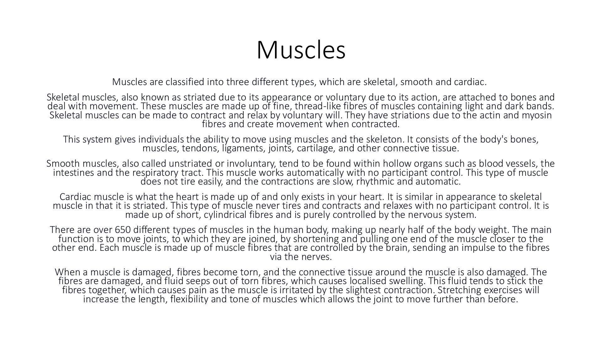
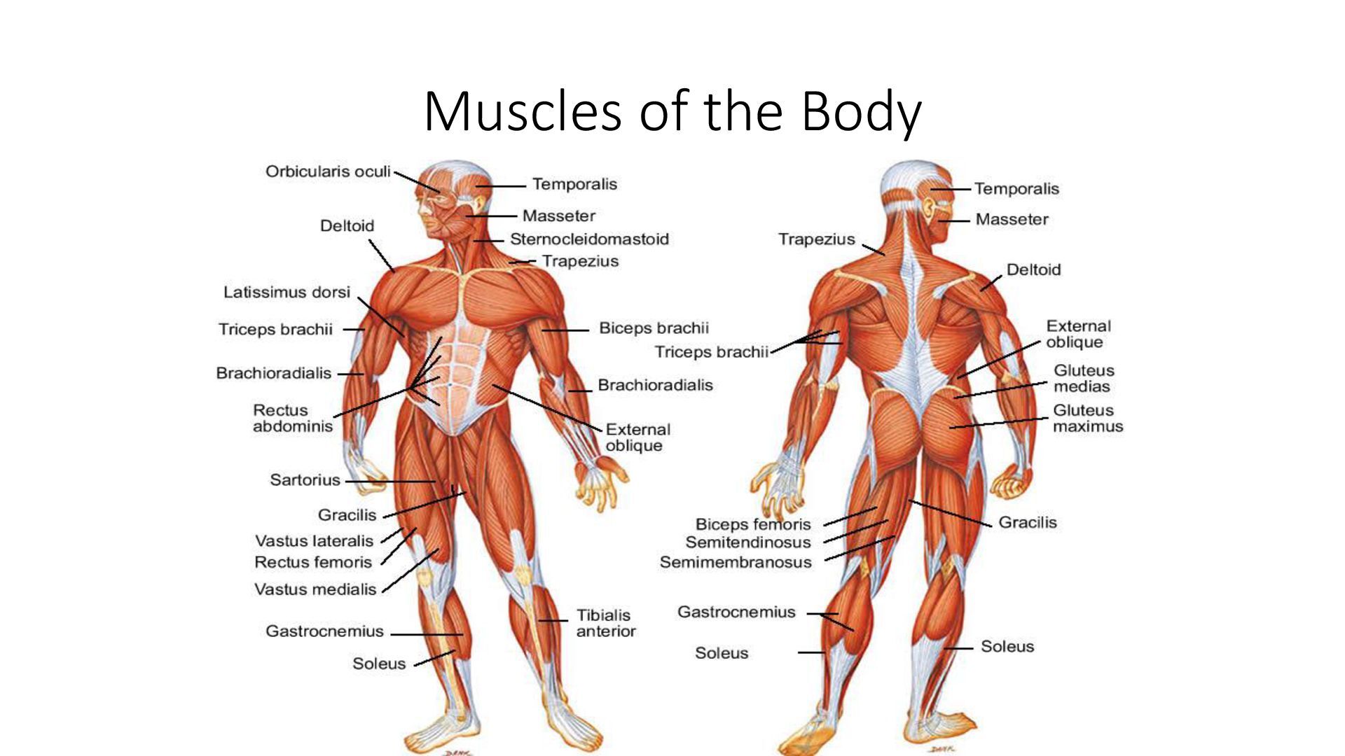
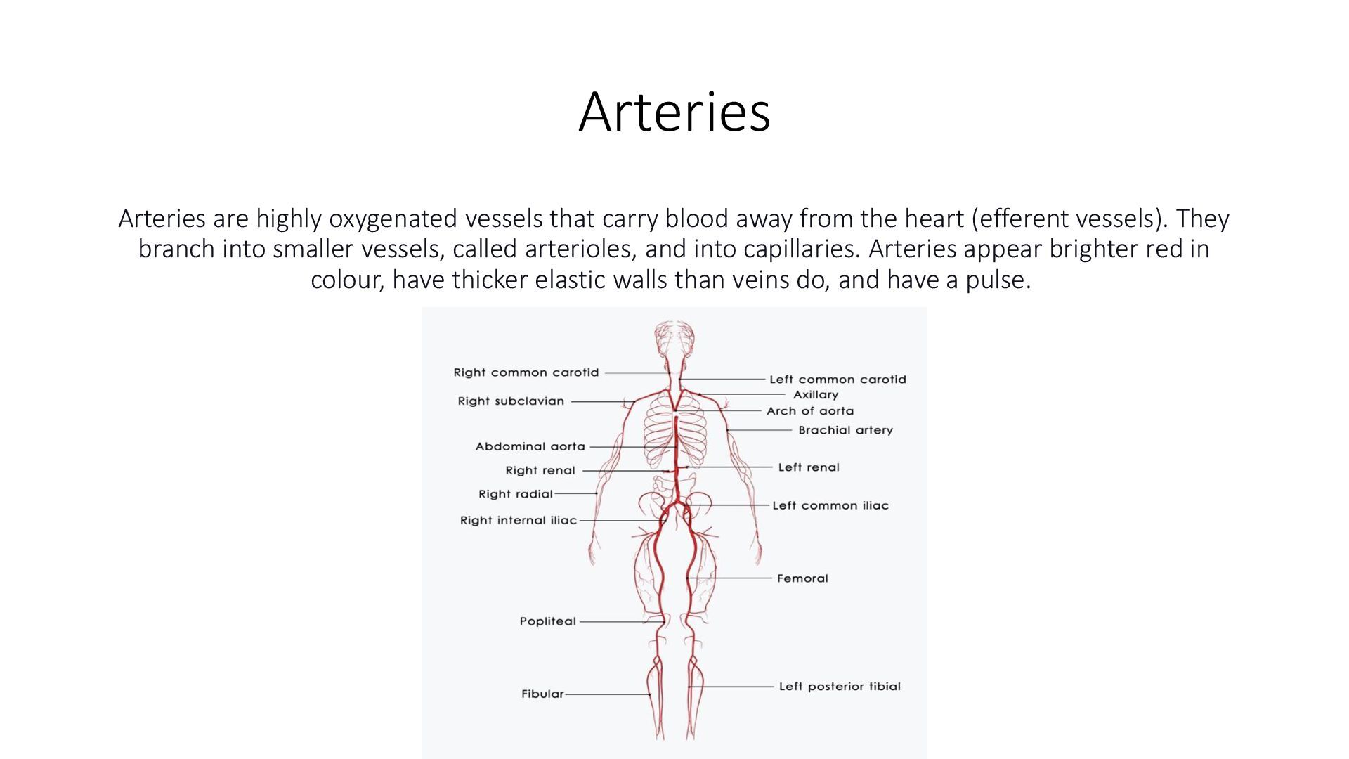
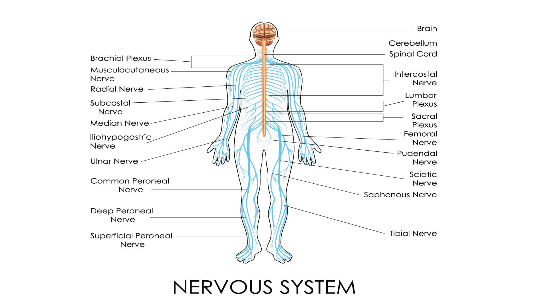
    human body, most of which are in the brain. The nervous system can be thought of as two systems: The central nervous system (CNS) is made up of the brain and spinal cord, and the peripheral nervous system (PNS) is everything outside of the brain and spinal cord. Sensory neurons transmit nerve impulses to the spinal cord or the brain from muscle tissues. Motor neurons transmit impulses to muscles from the spinal cord or the brain. Both the brain and the spinal cord are covered by protective membranes (meninges). Between these protective membrane layers are spaces filled with cerebrospinal fluid (CSF) that provides a cushion for the brain and the spinal cord. Furthermore, the brain and spinal cord are protected by the skull and vertebral column, respectively. The bony segments of the vertebral canal are divided into regions (cervical, thoracic, and lumbar vertebrae). There are seven cervical vertebrae (C1- C7) that extend from the head to the thorax, 12 thoracic vertebrae (T1 T12) that extend from the chest to the back, and five lumbar vertebrae (L1-L5) that extend to the lower back. At the lower end of the vertebral column, the sacrum (S1-S5) and coccyx are fused elements of the sacral and coccygeal vertebrae. The brain, along with the cranial nerves, functions in all mental processes and many essential motor, sensory, and visceral responses. The spinal cord and the spinal nerves control sensory (touch), motor (voluntary movement), and reflex (knee-jerk) functions. Reflexes are responses to stimuli that do not require communication with the brain. A simple reflex, such as moving a finger from something hot, occurs even before the brain realises the pain. Specific cranial and spinal nerves control all complex or simple action processes in the body. There are 31 pairs of spinal nerves (nerves that branch off the spinal cord), each of which is identified by its location to the nearest vertebrae. Nerves that branch from the spinal cord (C5 through Tl) and extend into the arm region (the brachial plexus) are the axillary, radial, musculocutaneous, median, and ulnar nerves. These nerves control all muscle movement of the shoulder, arm, and hand and also control sensations of the skin of the entire shoulder, arm, and hand. In summary, the nervous system is the primary communication and regulatory system in the body. The autonomic nervous system entails the functions that work without voluntary control of an individual, such as heartbeat, rate of breathing, tear and saliva production, and bladder constrictions.
  63. Disorders of the Nervous System ➢ Infectious conditions (viral or

    bacterial) such as encephalitis (inflammation of the brain), meningitis (inflammation of the linings of the brain and spinal cord), tetanus, herpes, and poliomyelitis. ➢ Conditions such as ALS, MS, Parkinson's disease (a degenerative disorder characterised by hand tremors, loss of facial expression, shuffling walk), cerebral palsy (CP; brain damage at birth that typically causes lack of muscle control) (FIGURE 6- 21) ➢ Epilepsy (episodes of abnormal electrical discharges in the brain that may cause convulsions or loss of consciousness), hydrocephaly (excessive amounts of CSF in the brain that can lead to intracranial pressure and other complications), neuralgia (pain along a nerve), strokes, and headaches ➢ Injuries that can also result in paralysis or partial paralysis • As a special note to those performing blood collection, nerve damage, including partial paralysis, can also occur as a result of accidental injury during phlebotomy procedures. Injury may be the result of excessive probing with the needle, sticking the needle in a poor site for venipuncture, deep needle penetration all the way into the nerve, and/or if the patient suddenly jerks his or her arm during the venipuncture procedure, causing the needle to puncture a nerve. Choose venipuncture sites that are stabilised as much as possible. Under no circumstances should you use the anterior or palmar side of the wrist to collect a blood specimen because the risk of hitting a nerve is high due to nerve locations close to the skin's surface.