Upgrade to Pro — share decks privately, control downloads, hide ads and more …

INTRODUCTION TO THE CELL

INTRODUCTION TO THE CELL

A cell is the fundamental structural, functional, and biological unit of all living organisms. Often referred to as the "building blocks of life," cells are the smallest entities capable of executing all life processes independently—from metabolizing nutrients to replicating genetic material.

Avatar for Pawan Kumar Sahu

Pawan Kumar Sahu

September 02, 2026

More Decks by Pawan Kumar Sahu

Other Decks in Education

Transcript

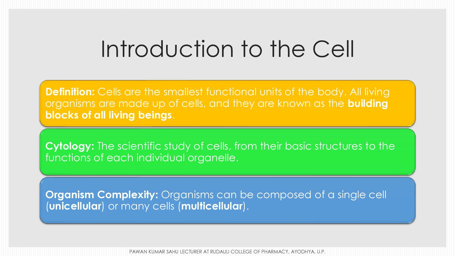
  1. Introduction to the Cell Definition: Cells are the smallest functional

    units of the body. All living organisms are made up of cells, and they are known as the building blocks of all living beings. Cytology: The scientific study of cells, from their basic structures to the functions of each individual organelle. Organism Complexity: Organisms can be composed of a single cell (unicellular) or many cells (multicellular). PAWAN KUMAR SAHU LECTURER AT RUDAULI COLLEGE OF PHARMACY, AYODHYA, U.P.
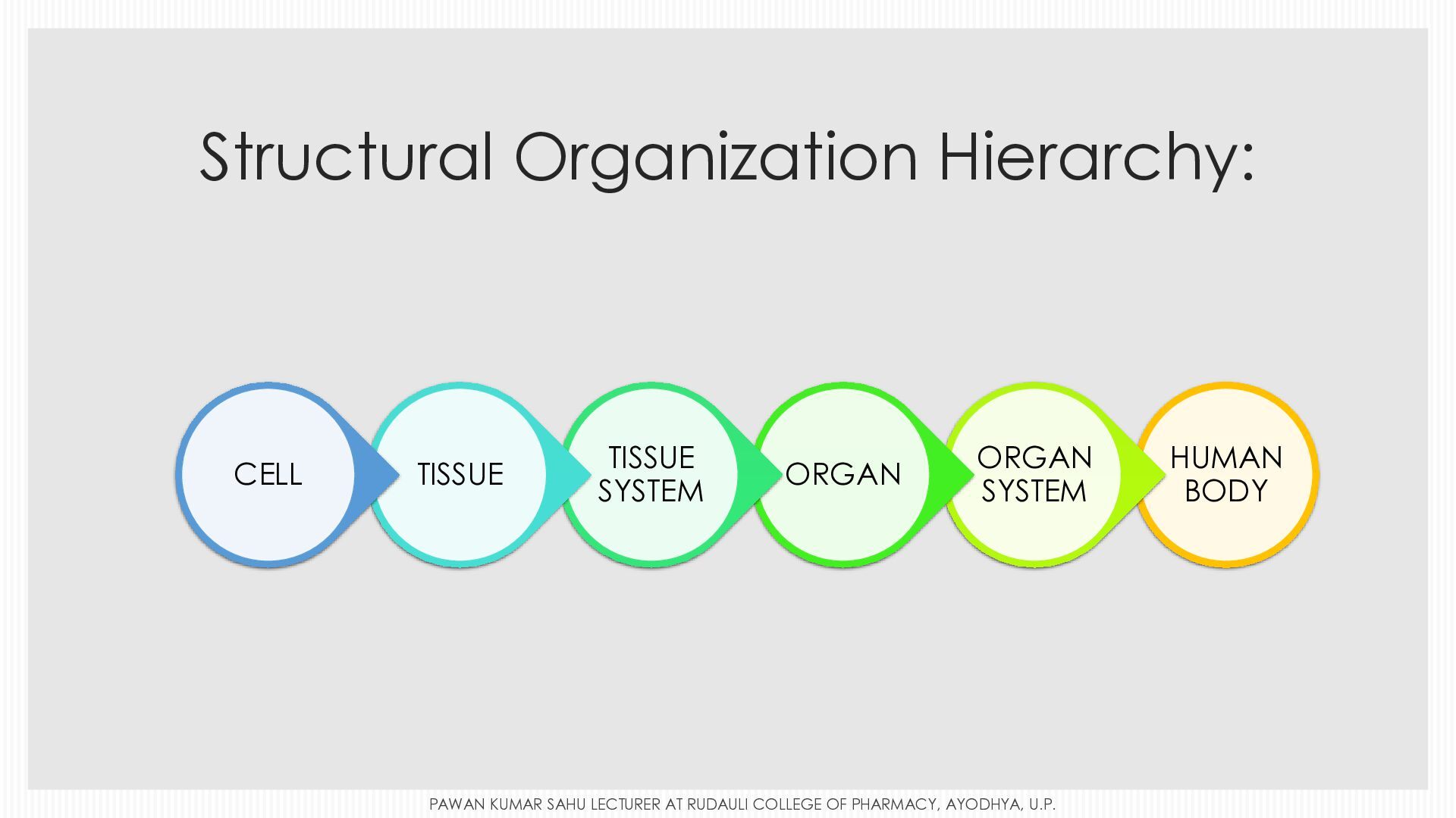
  2. Structural Organization Hierarchy: CELL TISSUE TISSUE SYSTEM ORGAN ORGAN SYSTEM

    PAWAN KUMAR SAHU LECTURER AT RUDAULI COLLEGE OF PHARMACY, AYODHYA, U.P. HUMAN BODY
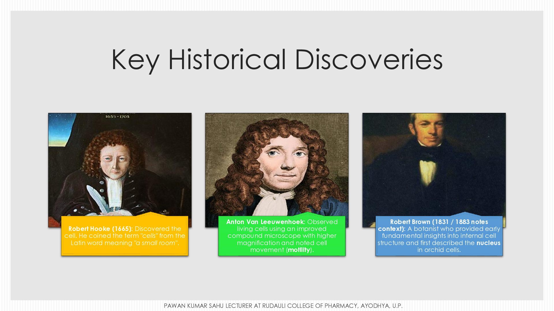
  3. Key Historical Discoveries Robert Hooke (1665): Discovered the cell. He

    coined the term "cells" from the Latin word meaning "a small room". Anton Van Leeuwenhoek: Observed living cells using an improved compound microscope with higher magnification and noted cell movement (motility). Robert Brown (1831 / 1883 notes context): A botanist who provided early fundamental insights into internal cell structure and first described the nucleus in orchid cells. PAWAN KUMAR SAHU LECTURER AT RUDAULI COLLEGE OF PHARMACY, AYODHYA, U.P.
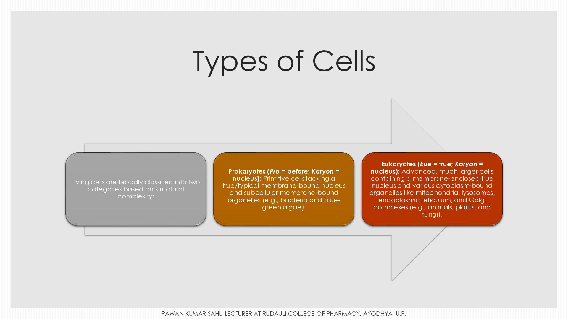
  4. Types of Cells Living cells are broadly classified into two

    categories based on structural complexity: Prokaryotes (Pro = before; Karyon = nucleus): Primitive cells lacking a true/typical membrane-bound nucleus and subcellular membrane-bound organelles (e.g., bacteria and bluegreen algae). Eukaryotes (Eue = true; Karyon = nucleus): Advanced, much larger cells containing a membrane-enclosed true nucleus and various cytoplasm-bound organelles like mitochondria, lysosomes, endoplasmic reticulum, and Golgi complexes (e.g., animals, plants, and fungi). PAWAN KUMAR SAHU LECTURER AT RUDAULI COLLEGE OF PHARMACY, AYODHYA, U.P.
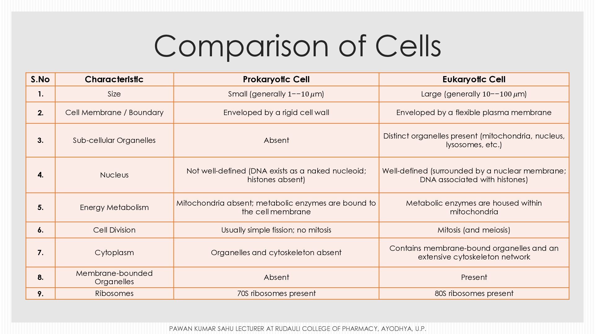
  5. Comparison of Cells S.No Characteristic Prokaryotic Cell Eukaryotic Cell 1.

    Size Small (generally 1−−10 𝜇m) Large (generally 10−−100 𝜇m) 2. Cell Membrane / Boundary Enveloped by a rigid cell wall Enveloped by a flexible plasma membrane 3. Sub-cellular Organelles Absent Distinct organelles present (mitochondria, nucleus, lysosomes, etc.) 4. Nucleus Not well-defined (DNA exists as a naked nucleoid; histones absent) Well-defined (surrounded by a nuclear membrane; DNA associated with histones) 5. Energy Metabolism Mitochondria absent; metabolic enzymes are bound to the cell membrane Metabolic enzymes are housed within mitochondria 6. Cell Division Usually simple fission; no mitosis Mitosis (and meiosis) 7. Cytoplasm Organelles and cytoskeleton absent Contains membrane-bound organelles and an extensive cytoskeleton network 8. Membrane-bounded Organelles Absent Present 9. Ribosomes 70S ribosomes present 80S ribosomes present PAWAN KUMAR SAHU LECTURER AT RUDAULI COLLEGE OF PHARMACY, AYODHYA, U.P.
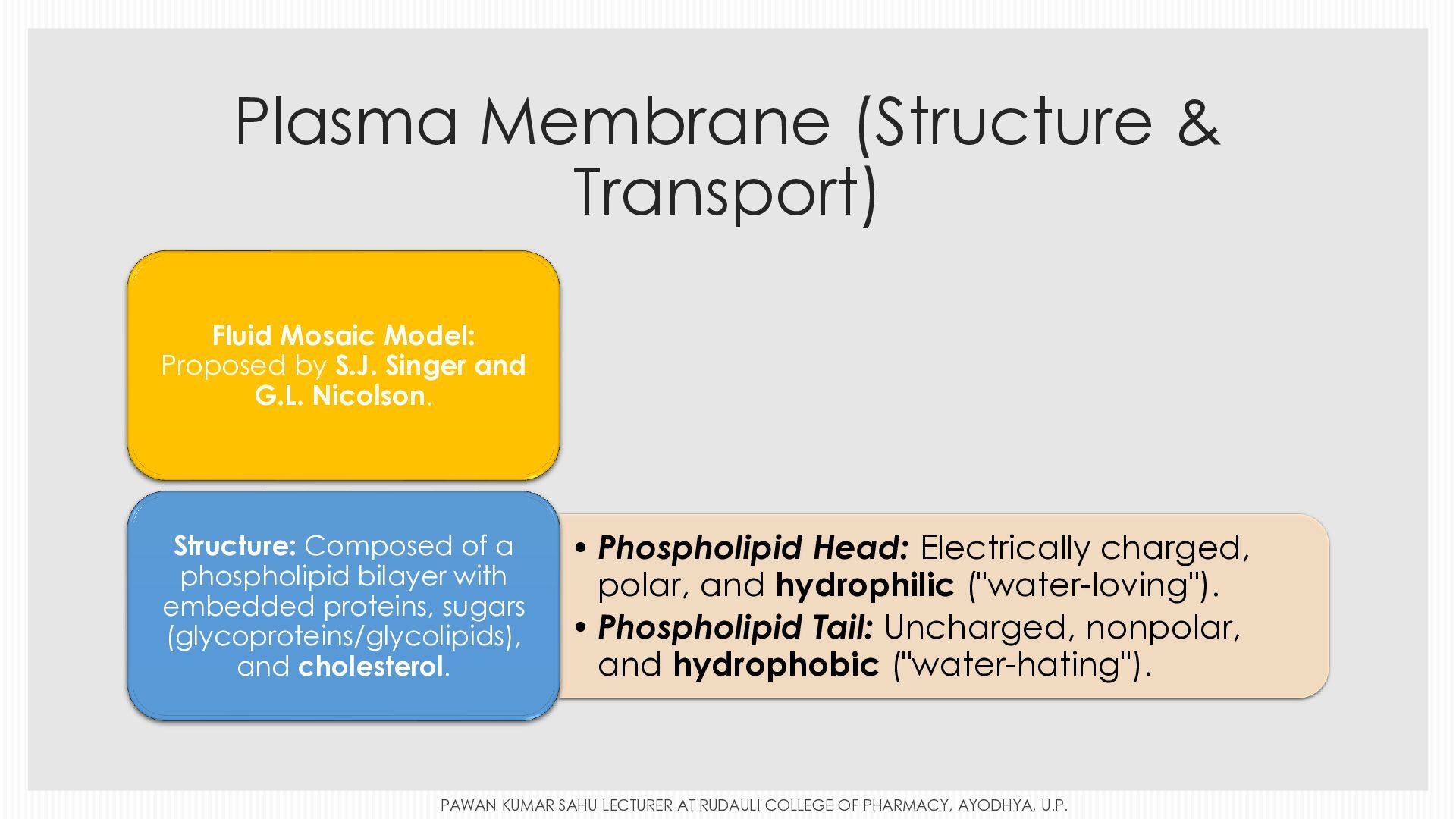
  6. Plasma Membrane (Structure & Transport) Fluid Mosaic Model: Proposed by

    S.J. Singer and G.L. Nicolson. Structure: Composed of a phospholipid bilayer with embedded proteins, sugars (glycoproteins/glycolipids), and cholesterol. • Phospholipid Head: Electrically charged, polar, and hydrophilic ("water-loving"). • Phospholipid Tail: Uncharged, nonpolar, and hydrophobic ("water-hating"). PAWAN KUMAR SAHU LECTURER AT RUDAULI COLLEGE OF PHARMACY, AYODHYA, U.P.
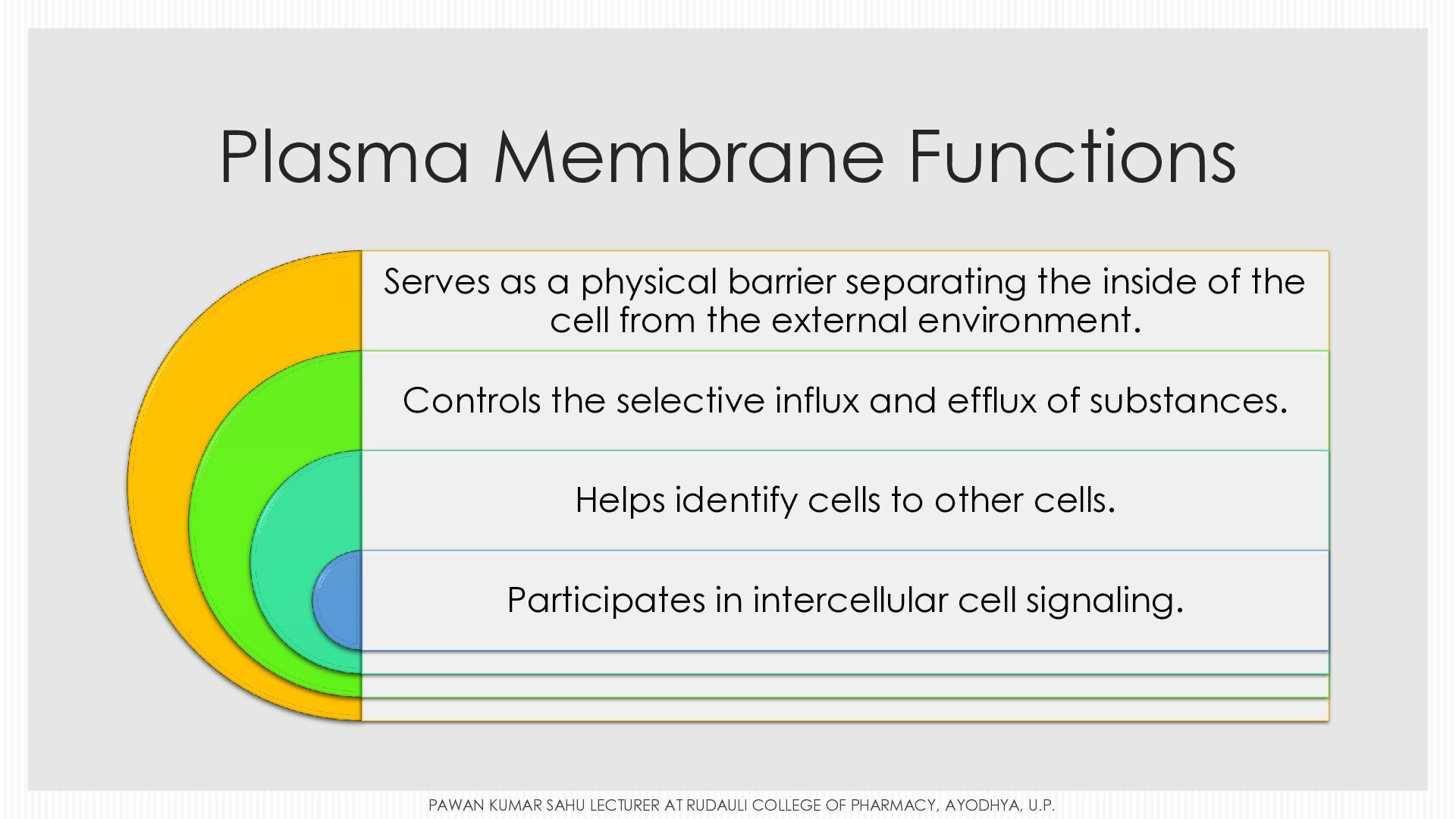
  7. Plasma Membrane Functions Serves as a physical barrier separating the

    inside of the cell from the external environment. Controls the selective influx and efflux of substances. Helps identify cells to other cells. Participates in intercellular cell signaling. PAWAN KUMAR SAHU LECTURER AT RUDAULI COLLEGE OF PHARMACY, AYODHYA, U.P.
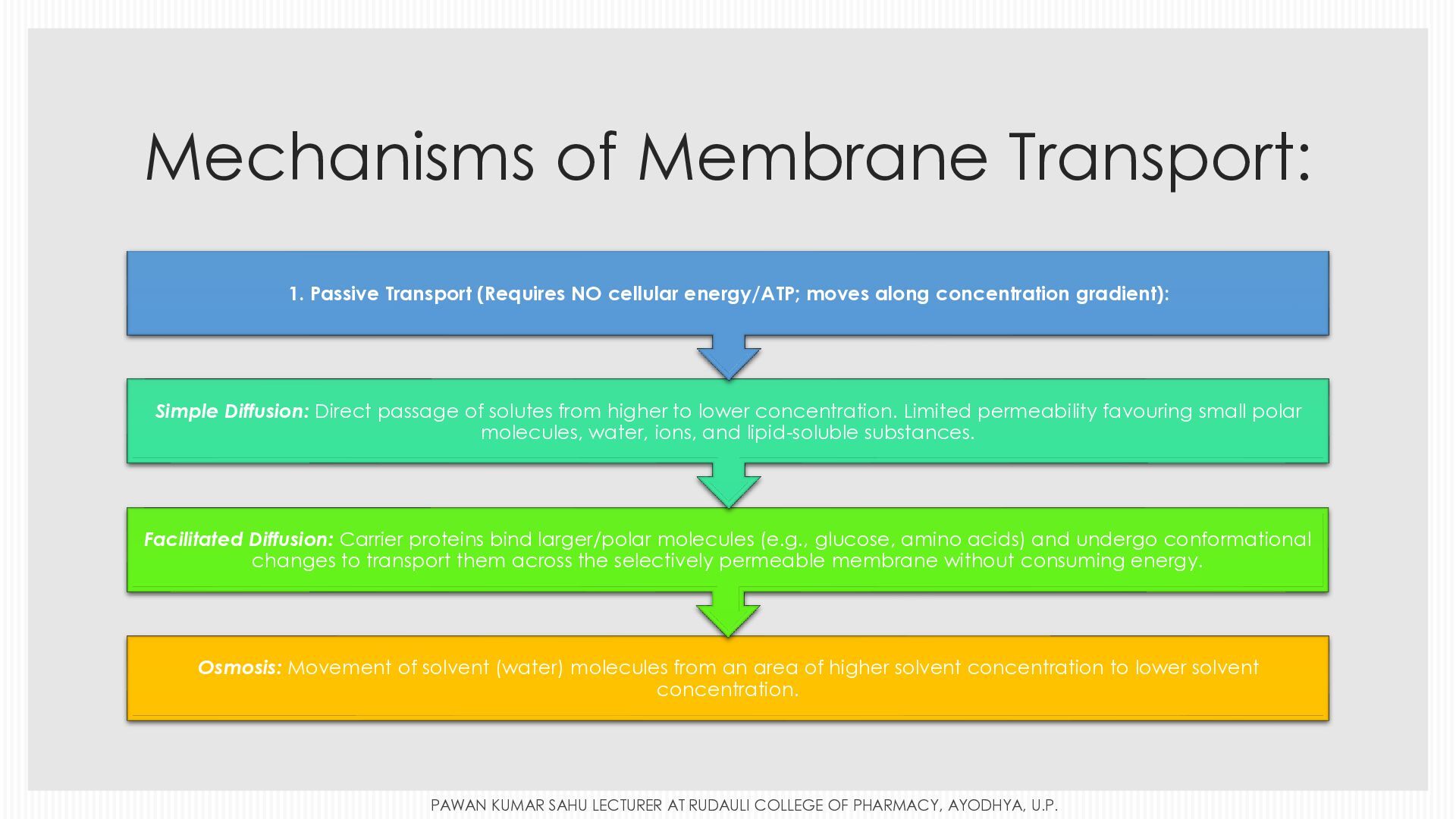
  8. Mechanisms of Membrane Transport: 1. Passive Transport (Requires NO cellular

    energy/ATP; moves along concentration gradient): Simple Diffusion: Direct passage of solutes from higher to lower concentration. Limited permeability favouring small polar molecules, water, ions, and lipid-soluble substances. Facilitated Diffusion: Carrier proteins bind larger/polar molecules (e.g., glucose, amino acids) and undergo conformational changes to transport them across the selectively permeable membrane without consuming energy. Osmosis: Movement of solvent (water) molecules from an area of higher solvent concentration to lower solvent concentration. PAWAN KUMAR SAHU LECTURER AT RUDAULI COLLEGE OF PHARMACY, AYODHYA, U.P.
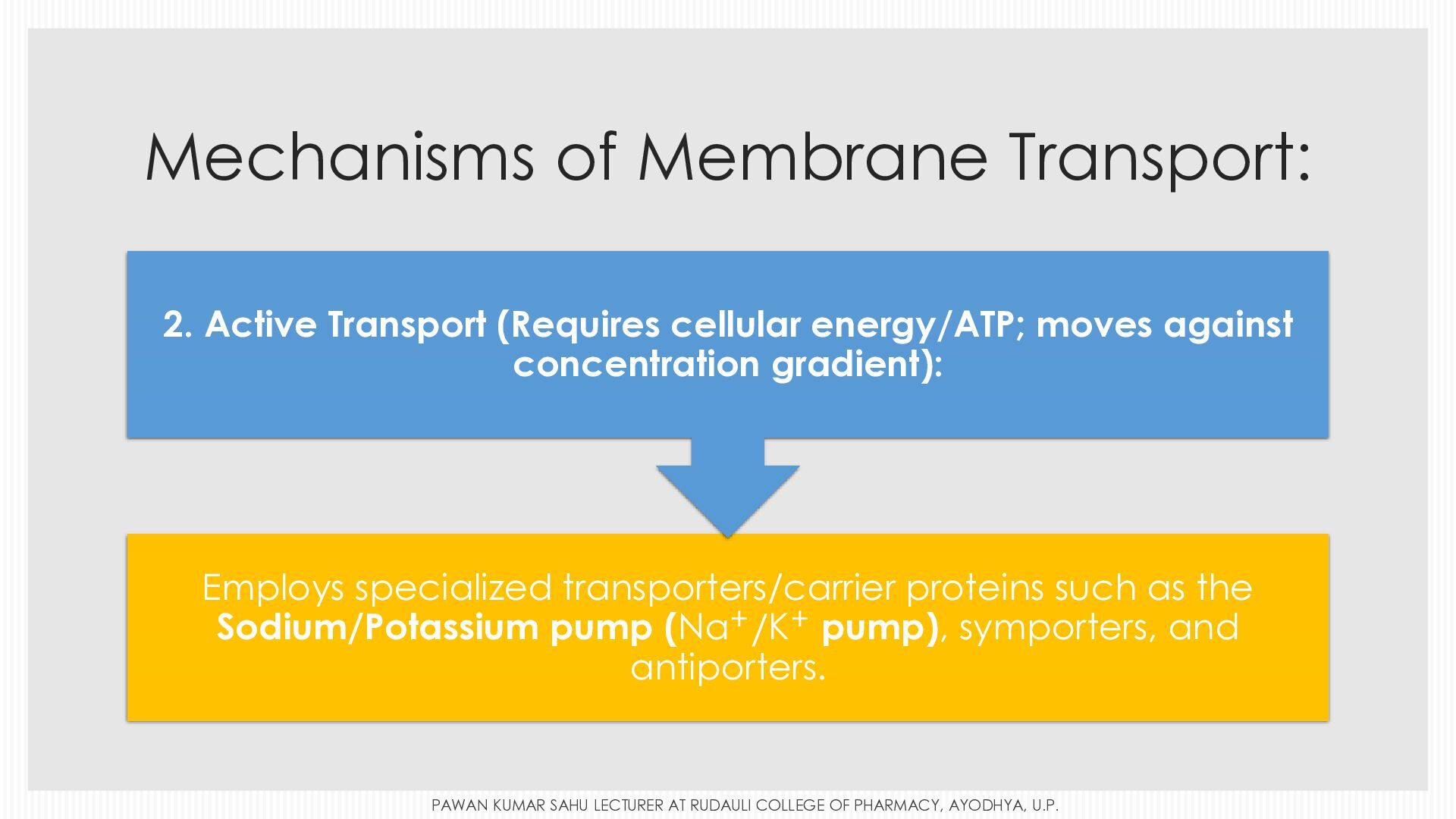
  9. Mechanisms of Membrane Transport: 2. Active Transport (Requires cellular energy/ATP;

    moves against concentration gradient): Employs specialized transporters/carrier proteins such as the Sodium/Potassium pump (Na+ /K+ pump), symporters, and antiporters. superscript base , Na , end base , to the plus over K to the plus PAWAN KUMAR SAHU LECTURER AT RUDAULI COLLEGE OF PHARMACY, AYODHYA, U.P.
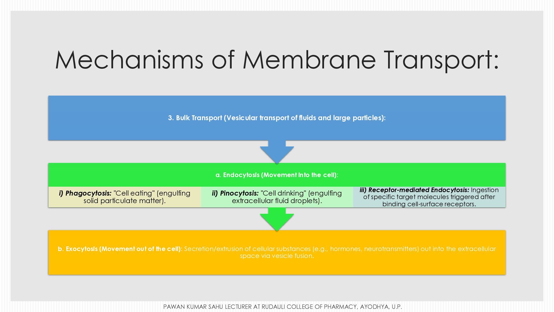
  10. Mechanisms of Membrane Transport: 3. Bulk Transport (Vesicular transport of

    fluids and large particles): a. Endocytosis (Movement into the cell): i) Phagocytosis: "Cell eating" (engulfing solid particulate matter). ii) Pinocytosis: "Cell drinking" (engulfing extracellular fluid droplets). iii) Receptor-mediated Endocytosis: Ingestion of specific target molecules triggered after binding cell-surface receptors. b. Exocytosis (Movement out of the cell): Secretion/extrusion of cellular substances (e.g., hormones, neurotransmitters) out into the extracellular space via vesicle fusion. PAWAN KUMAR SAHU LECTURER AT RUDAULI COLLEGE OF PHARMACY, AYODHYA, U.P.
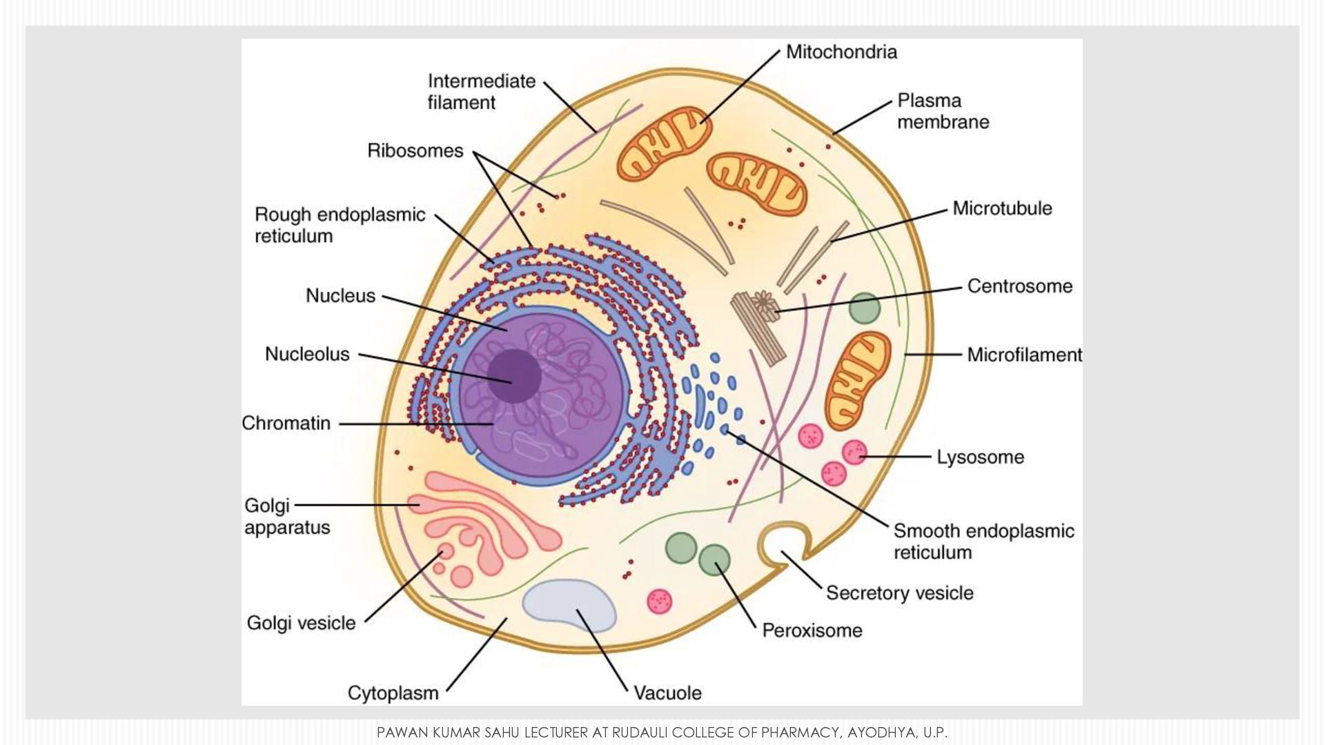
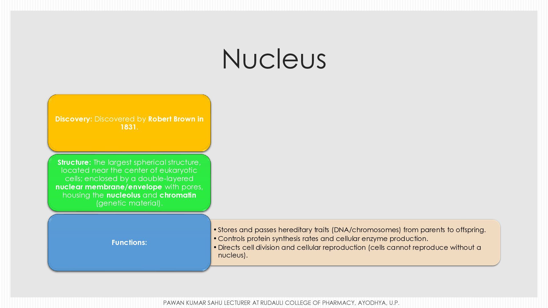
  11. Nucleus Discovery: Discovered by Robert Brown in 1831. Structure: The

    largest spherical structure, located near the center of eukaryotic cells; enclosed by a double-layered nuclear membrane/envelope with pores, housing the nucleolus and chromatin (genetic material). Functions: • Stores and passes hereditary traits (DNA/chromosomes) from parents to offspring. • Controls protein synthesis rates and cellular enzyme production. • Directs cell division and cellular reproduction (cells cannot reproduce without a nucleus). PAWAN KUMAR SAHU LECTURER AT RUDAULI COLLEGE OF PHARMACY, AYODHYA, U.P.
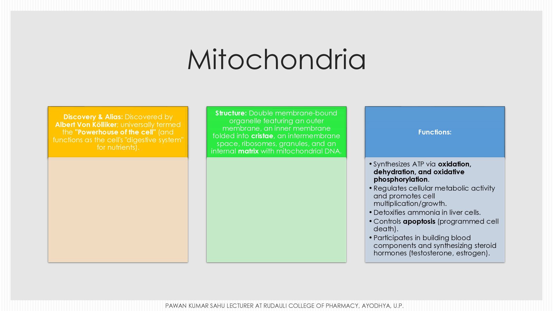
  12. Mitochondria Discovery & Alias: Discovered by Albert Von Kölliker; universally

    termed the "Powerhouse of the cell" (and functions as the cell's "digestive system" for nutrients). Structure: Double membrane-bound organelle featuring an outer membrane, an inner membrane folded into cristae, an intermembrane space, ribosomes, granules, and an internal matrix with mitochondrial DNA. Functions: • Synthesizes ATP via oxidation, dehydration, and oxidative phosphorylation. • Regulates cellular metabolic activity and promotes cell multiplication/growth. • Detoxifies ammonia in liver cells. • Controls apoptosis (programmed cell death). • Participates in building blood components and synthesizing steroid hormones (testosterone, estrogen). PAWAN KUMAR SAHU LECTURER AT RUDAULI COLLEGE OF PHARMACY, AYODHYA, U.P.
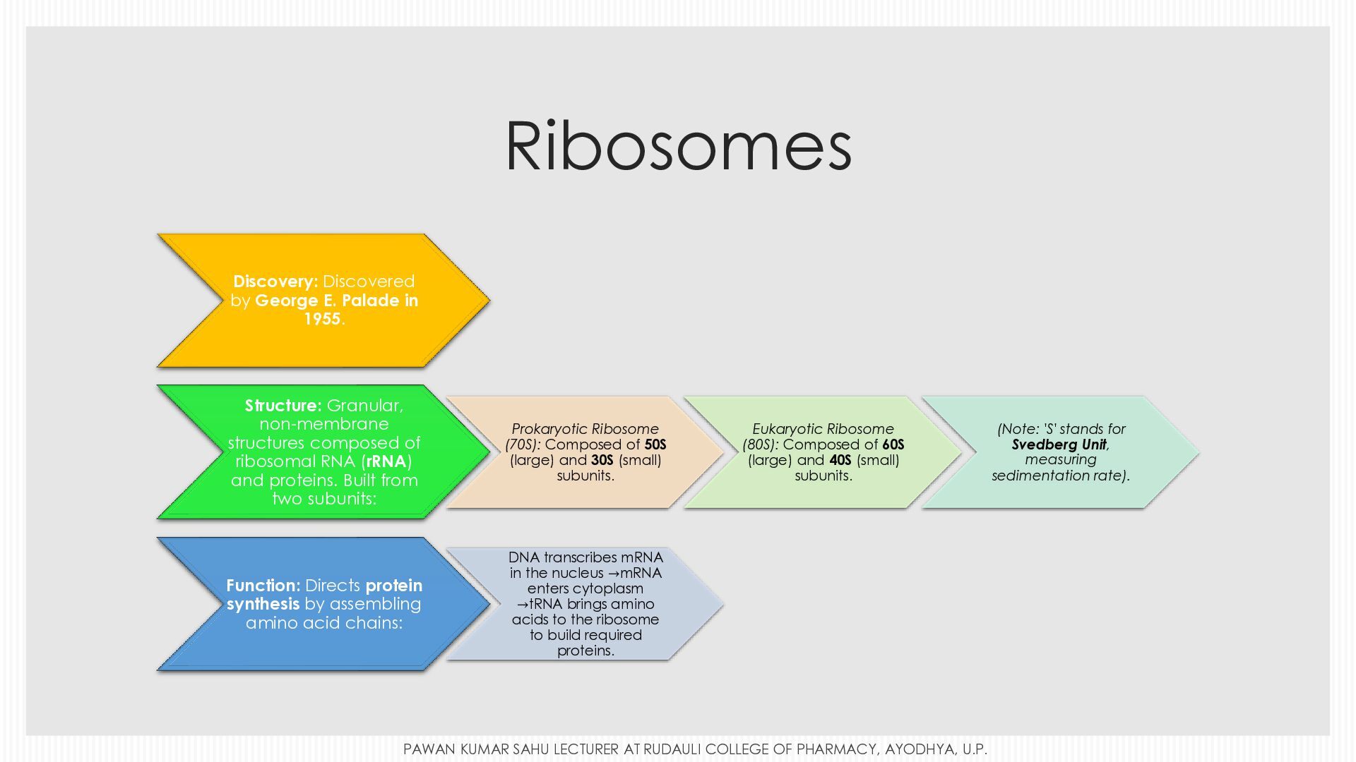
  13. Ribosomes Discovery: Discovered by George E. Palade in 1955. Structure:

    Granular, non-membrane structures composed of ribosomal RNA (rRNA) and proteins. Built from two subunits: Prokaryotic Ribosome (70S): Composed of 50S (large) and 30S (small) subunits. Function: Directs protein synthesis by assembling amino acid chains: DNA transcribes mRNA in the nucleus →mRNA enters cytoplasm →tRNA brings amino acids to the ribosome to build required proteins. Eukaryotic Ribosome (80S): Composed of 60S (large) and 40S (small) subunits. start equation goes to start equation goes to PAWAN KUMAR SAHU LECTURER AT RUDAULI COLLEGE OF PHARMACY, AYODHYA, U.P. (Note: 'S' stands for Svedberg Unit, measuring sedimentation rate).
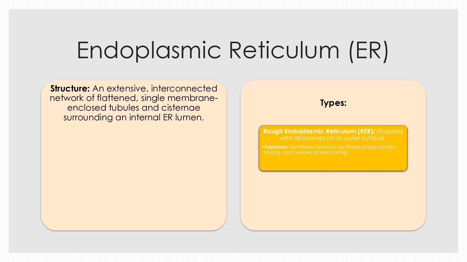
  14. Endoplasmic Reticulum (ER) Structure: An extensive, interconnected network of flattened,

    single membraneenclosed tubules and cisternae surrounding an internal ER lumen. Types: Rough Endoplasmic Reticulum (RER): Studded with ribosomes on its outer surface. •Functions: Synthesizes proteins, facilitates proper protein folding, and handles protein sorting.
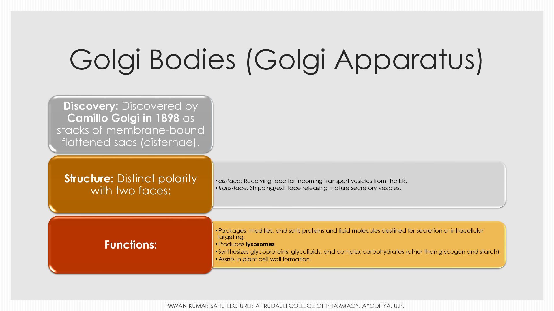
  15. Golgi Bodies (Golgi Apparatus) Discovery: Discovered by Camillo Golgi in

    1898 as stacks of membrane-bound flattened sacs (cisternae). Structure: Distinct polarity with two faces: Functions: •cis-face: Receiving face for incoming transport vesicles from the ER. •trans-face: Shipping/exit face releasing mature secretory vesicles. •Packages, modifies, and sorts proteins and lipid molecules destined for secretion or intracellular targeting. •Produces lysosomes. •Synthesizes glycoproteins, glycolipids, and complex carbohydrates (other than glycogen and starch). •Assists in plant cell wall formation. PAWAN KUMAR SAHU LECTURER AT RUDAULI COLLEGE OF PHARMACY, AYODHYA, U.P.
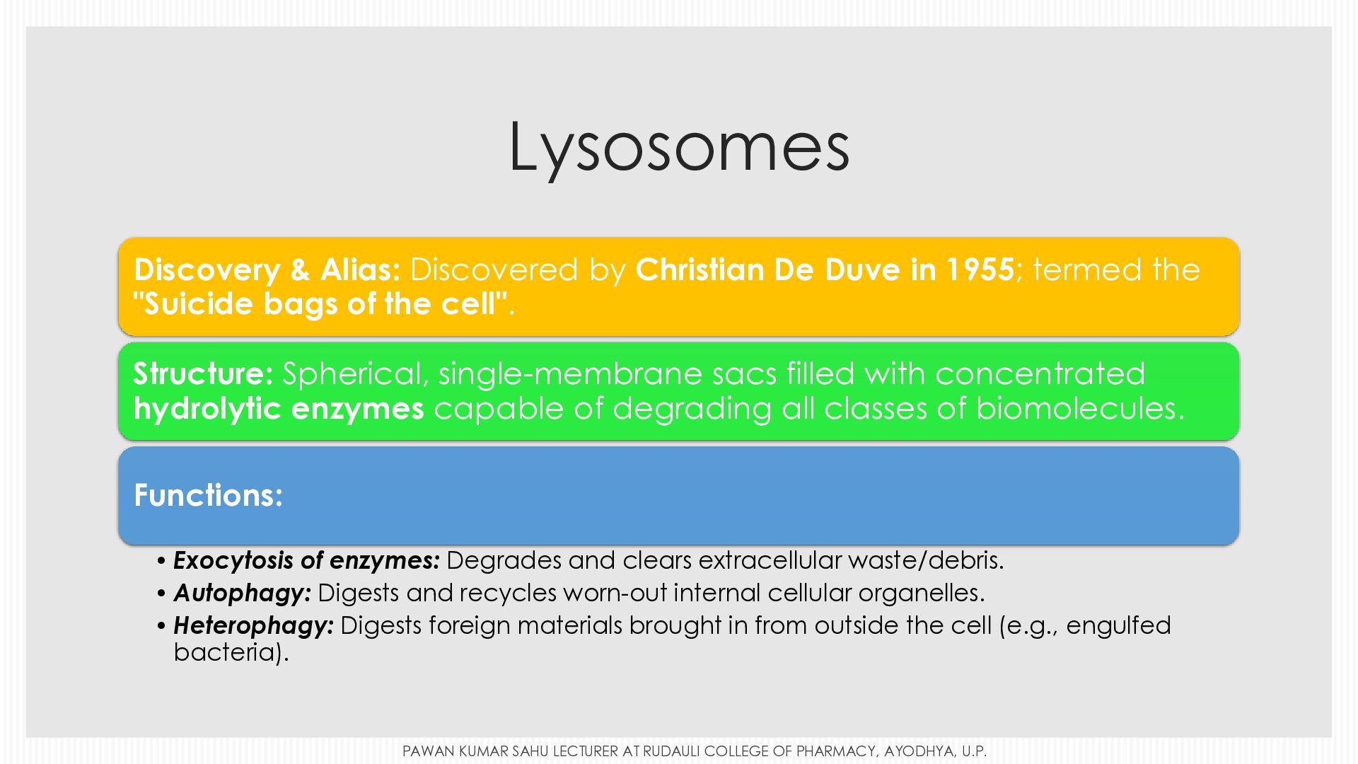
  16. Lysosomes Discovery & Alias: Discovered by Christian De Duve in

    1955; termed the "Suicide bags of the cell". Structure: Spherical, single-membrane sacs filled with concentrated hydrolytic enzymes capable of degrading all classes of biomolecules. Functions: • Exocytosis of enzymes: Degrades and clears extracellular waste/debris. • Autophagy: Digests and recycles worn-out internal cellular organelles. • Heterophagy: Digests foreign materials brought in from outside the cell (e.g., engulfed bacteria). PAWAN KUMAR SAHU LECTURER AT RUDAULI COLLEGE OF PHARMACY, AYODHYA, U.P.
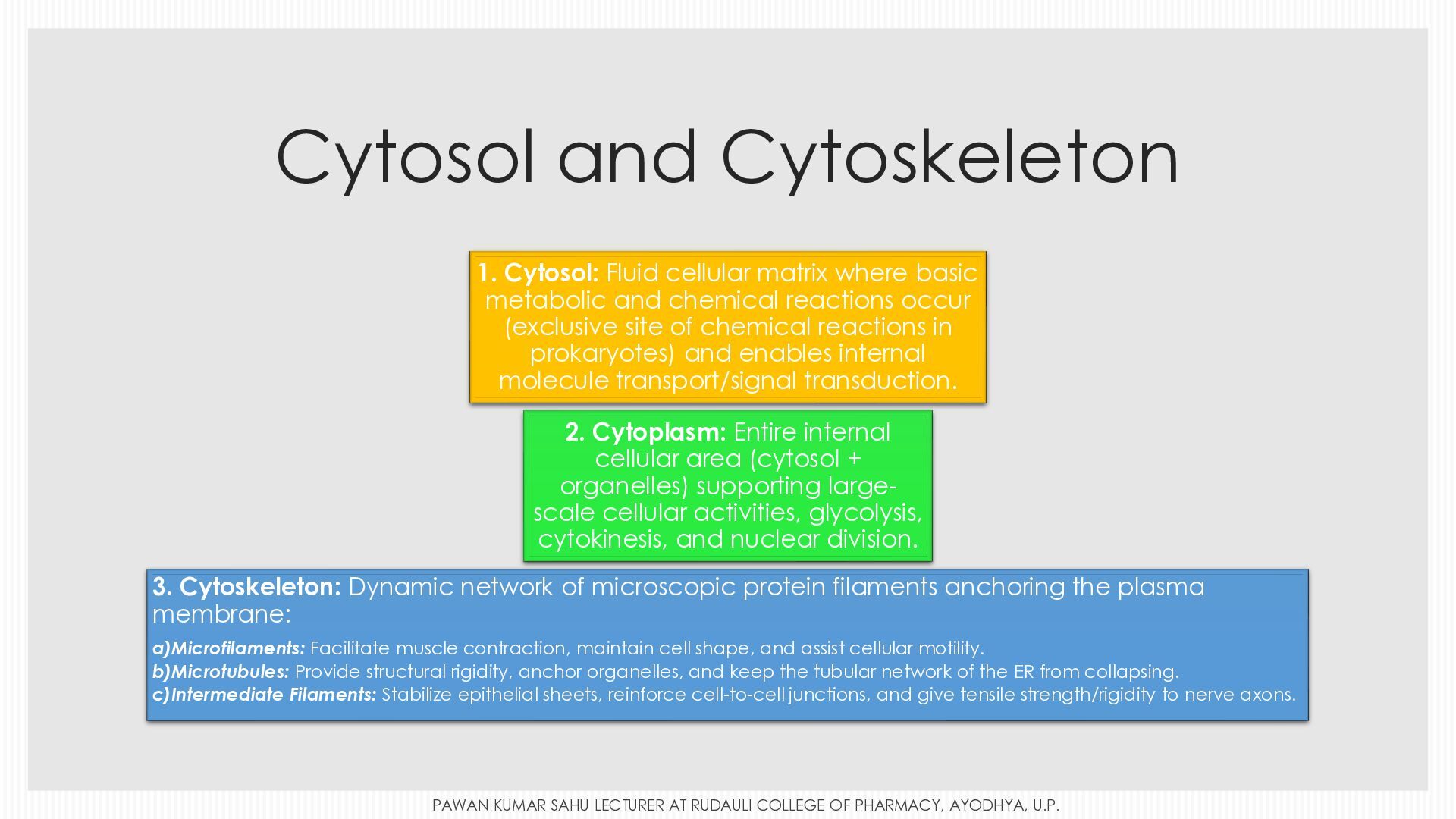
  17. Cytosol and Cytoskeleton 1. Cytosol: Fluid cellular matrix where basic

    metabolic and chemical reactions occur (exclusive site of chemical reactions in prokaryotes) and enables internal molecule transport/signal transduction. 2. Cytoplasm: Entire internal cellular area (cytosol + organelles) supporting largescale cellular activities, glycolysis, cytokinesis, and nuclear division. 3. Cytoskeleton: Dynamic network of microscopic protein filaments anchoring the plasma membrane: a)Microfilaments: Facilitate muscle contraction, maintain cell shape, and assist cellular motility. b)Microtubules: Provide structural rigidity, anchor organelles, and keep the tubular network of the ER from collapsing. c)Intermediate Filaments: Stabilize epithelial sheets, reinforce cell-to-cell junctions, and give tensile strength/rigidity to nerve axons. PAWAN KUMAR SAHU LECTURER AT RUDAULI COLLEGE OF PHARMACY, AYODHYA, U.P.
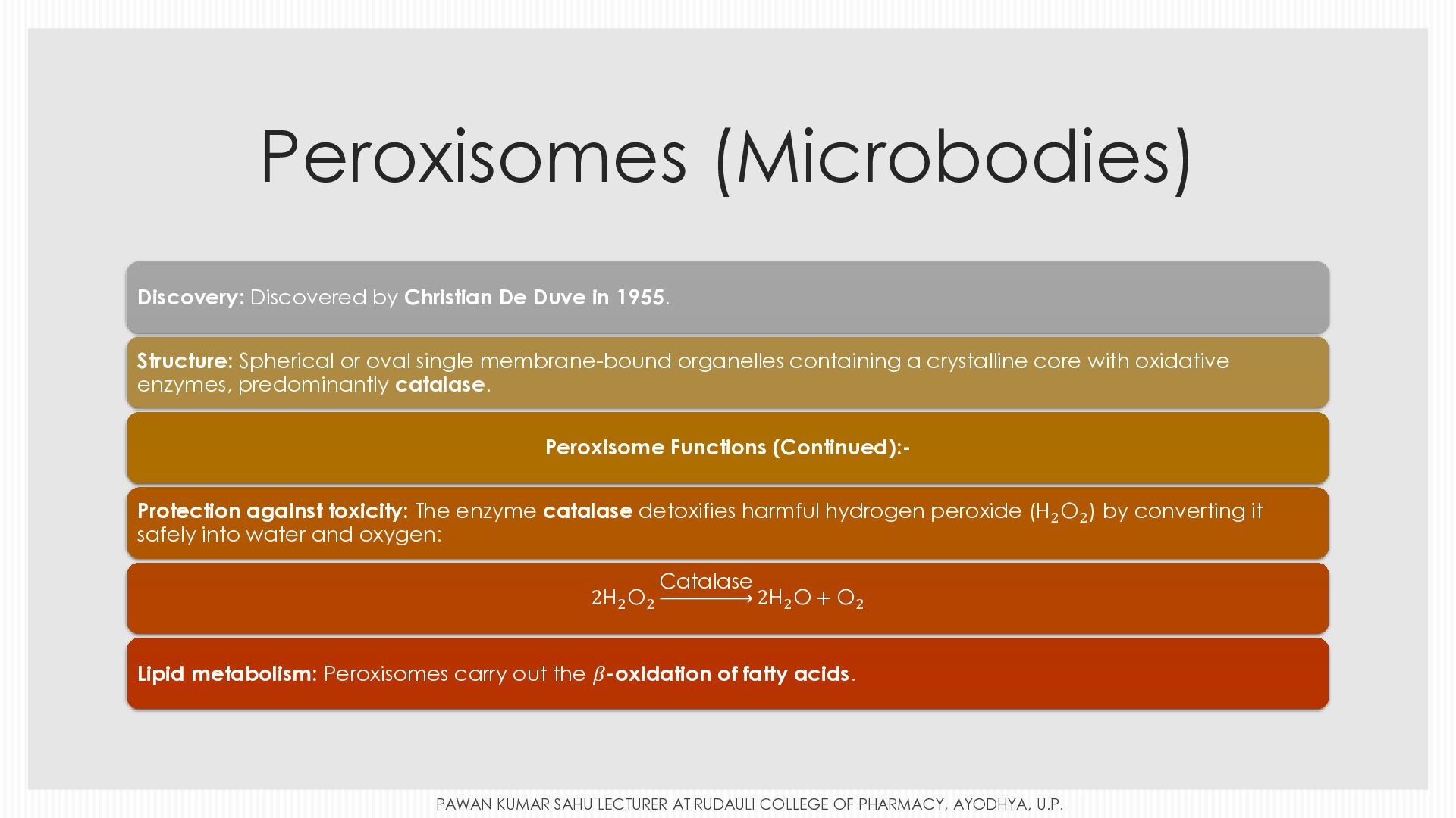
  18. Peroxisomes (Microbodies) Discovery: Discovered by Christian De Duve in 1955.

    Structure: Spherical or oval single membrane-bound organelles containing a crystalline core with oxidative enzymes, predominantly catalase. Peroxisome Functions (Continued):Protection against toxicity: The enzyme catalase detoxifies harmful hydrogen peroxide (H2 O2 ) by converting it safely into water and oxygen: H sub 2 , O sub 2 Catalase 2H sub 2 , O sub 2 , goes to upper limit □ open paren Catalase close paren , 2H sub 2 , O plus O sub 2 2H2 O2 2H2 O + O2 Lipid metabolism: Peroxisomes carry out the 𝛽-oxidation of fatty acids. start equation beta PAWAN KUMAR SAHU LECTURER AT RUDAULI COLLEGE OF PHARMACY, AYODHYA, U.P.
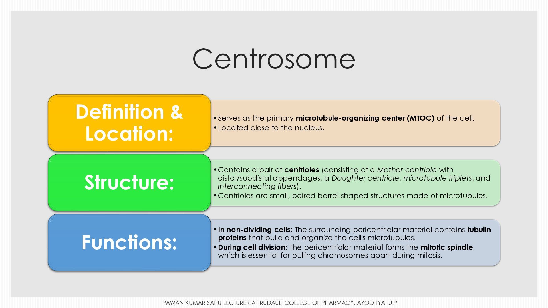
  19. Centrosome Definition & Location: •Serves as the primary microtubule-organizing center

    (MTOC) of the cell. •Located close to the nucleus. Structure: •Contains a pair of centrioles (consisting of a Mother centriole with distal/subdistal appendages, a Daughter centriole, microtubule triplets, and interconnecting fibers). •Centrioles are small, paired barrel-shaped structures made of microtubules. Functions: •In non-dividing cells: The surrounding pericentriolar material contains tubulin proteins that build and organize the cell's microtubules. •During cell division: The pericentriolar material forms the mitotic spindle, which is essential for pulling chromosomes apart during mitosis. PAWAN KUMAR SAHU LECTURER AT RUDAULI COLLEGE OF PHARMACY, AYODHYA, U.P.
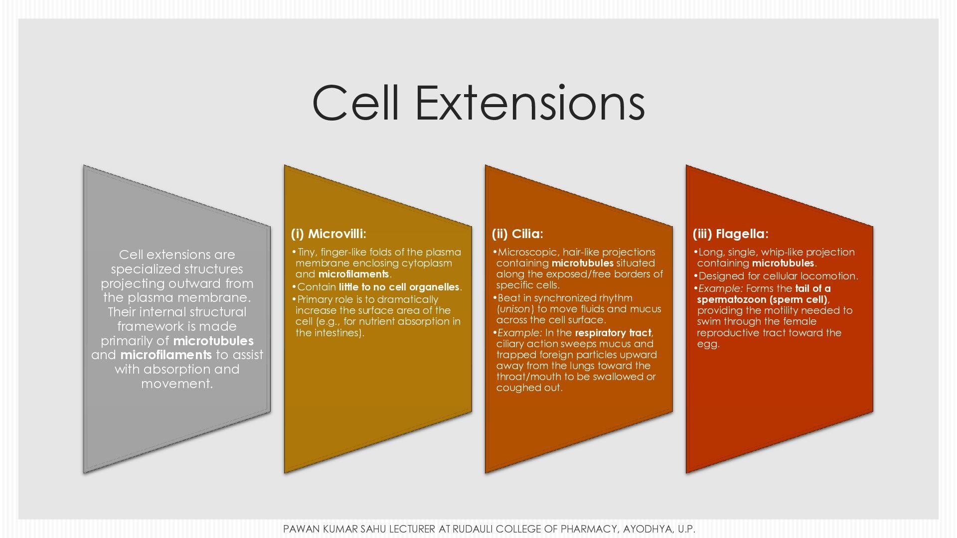
  20. Cell Extensions Cell extensions are specialized structures projecting outward from

    the plasma membrane. Their internal structural framework is made primarily of microtubules and microfilaments to assist with absorption and movement. (i) Microvilli: (ii) Cilia: (iii) Flagella: •Tiny, finger-like folds of the plasma membrane enclosing cytoplasm and microfilaments. •Contain little to no cell organelles. •Primary role is to dramatically increase the surface area of the cell (e.g., for nutrient absorption in the intestines). •Microscopic, hair-like projections containing microtubules situated along the exposed/free borders of specific cells. •Beat in synchronized rhythm (unison) to move fluids and mucus across the cell surface. •Example: In the respiratory tract, ciliary action sweeps mucus and trapped foreign particles upward away from the lungs toward the throat/mouth to be swallowed or coughed out. •Long, single, whip-like projection containing microtubules. •Designed for cellular locomotion. •Example: Forms the tail of a spermatozoon (sperm cell), providing the motility needed to swim through the female reproductive tract toward the egg. PAWAN KUMAR SAHU LECTURER AT RUDAULI COLLEGE OF PHARMACY, AYODHYA, U.P.