Upgrade to Pro — share decks privately, control downloads, hide ads and more …

TISSUE

 TISSUE

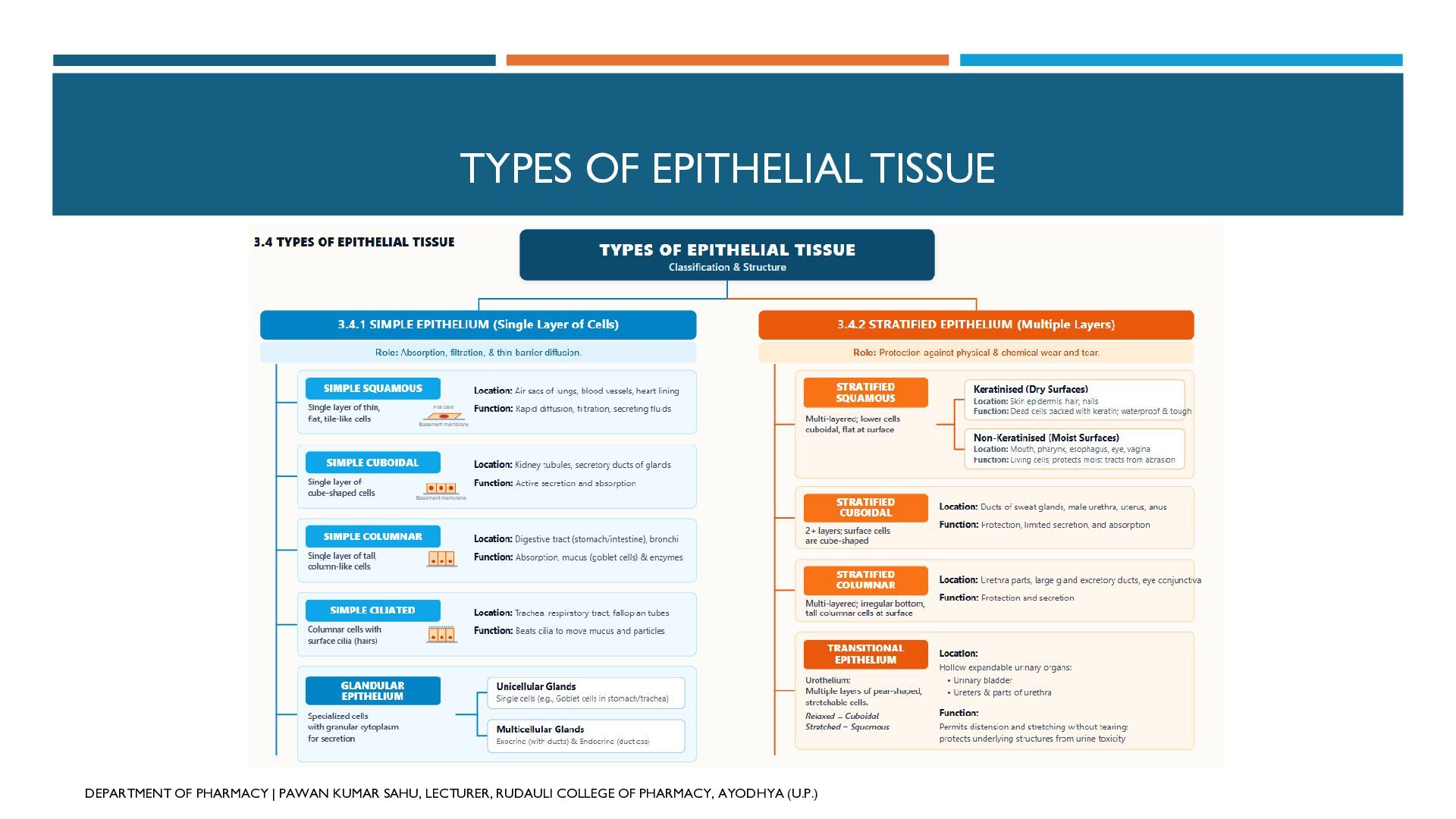
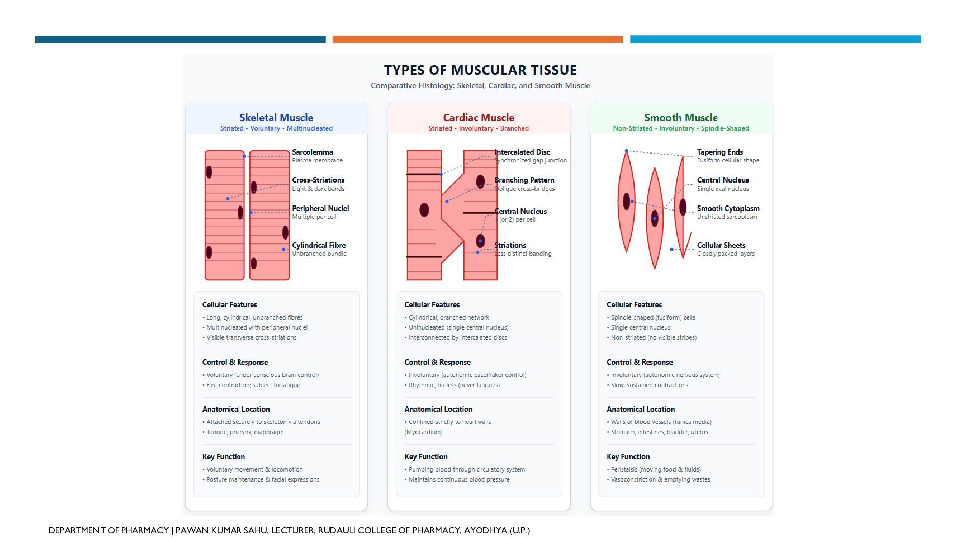
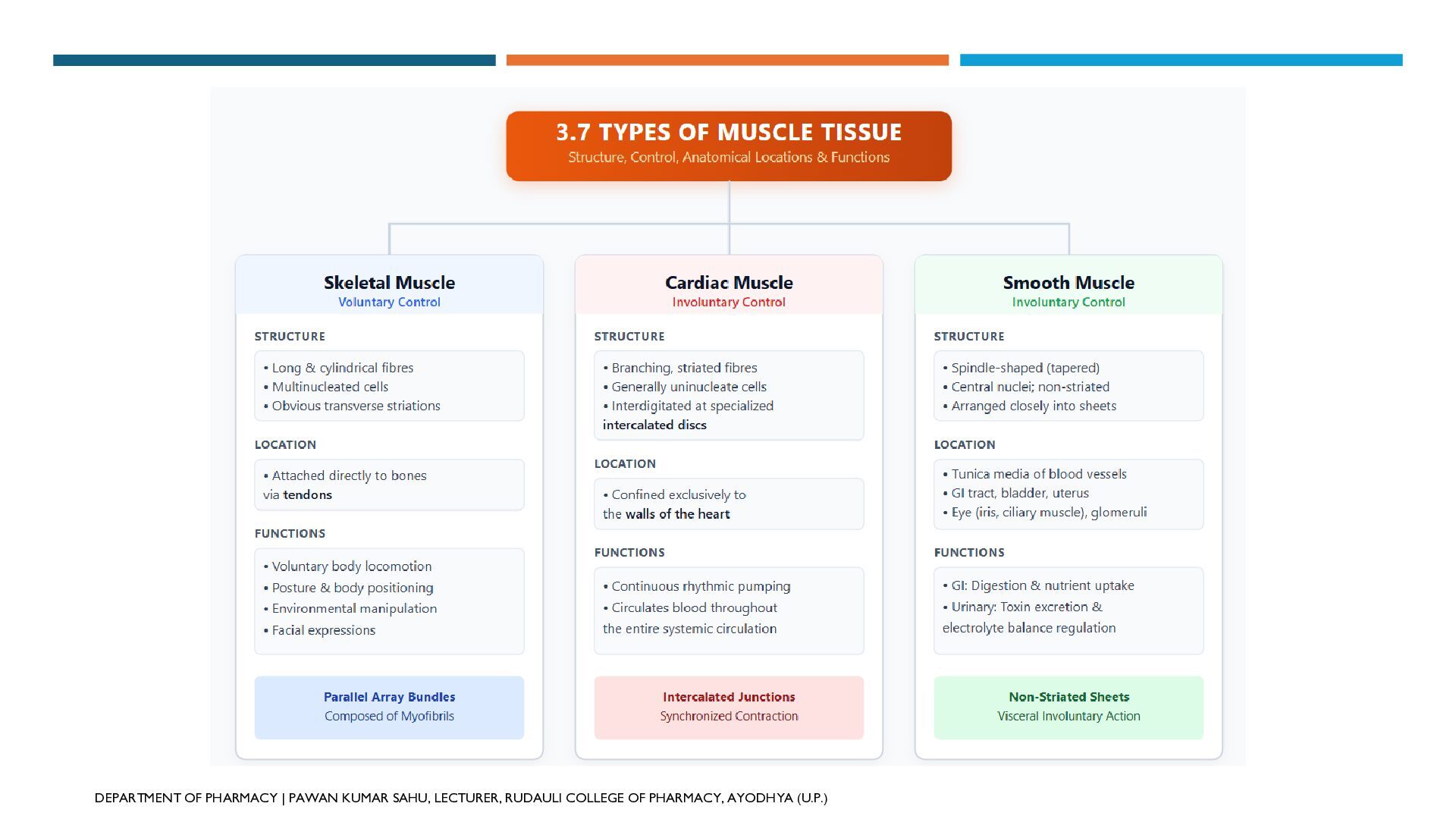
A tissue is a group of structurally similar cells and their intercellular substances that work together to perform specific biological functions. The human body comprises four primary tissue types:1. Epithelial TissueNature: Sheets of closely packed cells resting on a non-cellular basement membrane, with minimal intercellular matrix. Classification:Simple Epithelium (Single Layer): Specialized for absorption, secretion, and filtration. Subtypes include simple squamous (alveoli, endothelium), simple cuboidal (kidney tubules), simple columnar (stomach, intestines), simple ciliated (respiratory tract, fallopian tubes), and glandular epithelium (goblet cells). Stratified Epithelium (Multiple Layers): Specialized for mechanical and chemical protection against wear and tear. Subtypes include stratified squamous (keratinised in skin epidermis; non-keratinised in mouth and esophagus), stratified cuboidal (sweat gland ducts), stratified columnar (male urethra parts), and transitional epithelium/urothelium (urinary bladder, ureters). Primary Functions: Protection, selective absorption, secretion, excretion, and sensory reception. 2. Connective TissueNature: The most abundant and widely distributed tissue in complex animals. Consists of living cells scattered throughout an abundant non-living extracellular matrix containing protein fibres (collagen, elastic, reticular). Classification:Loose Connective Tissue: Fibres and cells are loosely woven in a semi-fluid ground substance. Includes areolar tissue (subcutaneous packing), adipose tissue (fat storage and thermal insulation), and reticular tissue (lymphoid organ stroma). Dense Connective Tissue: Densely packed fibres. Includes dense regular (tendons, ligaments), dense irregular (dermis of skin), and dense elastic (arterial walls, trachea). Specialized Skeletal Tissue: Firm and mineralized support tissues, namely cartilage (hyaline, fibrocartilage, elastic) and bone (calcified matrix with osteons/Haversian systems). Fluid Connective Tissue: Liquid matrix (plasma) with suspended cells, namely blood (erythrocytes, leukocytes, thrombocytes) and lymph. Primary Functions: Binds and supports organs, mechanical protection, thermal insulation, nutrient/waste exchange, and systemic transport. 3. Muscle TissueNature: Highly specialized contractile tissue made of elongated muscle cells (muscle fibres) containing contractile myofibrils. Classification:Skeletal Muscle: Long, cylindrical, multinucleated, striated fibres under voluntary control; attached to bones via tendons to drive body locomotion. Cardiac Muscle: Cylindrical, branched, uninucleated, striated fibres connected by intercalated discs; under involuntary control; confined to the heart wall (myocardium) to pump blood. Smooth Muscle: Spindle-shaped (fusiform), uninucleated, non-striated fibres arranged in sheets; under involuntary control; forms the walls of hollow organs (GI tract, blood vessels, bladder) to control peristalsis and vascular tone. Primary Functions: Body movement, posture maintenance, cardiac pumping, and involuntary visceral propulsion. 4. Nervous TissueNature: Specialized communication network dedicated to excitability and conduction. Fundamental Unit: The neuron (nerve cell), supported and insulated by neuroglial cells. Structure of a Neuron: Comprises a central cell body (soma) containing the nucleus, receiving branches (dendrites), and a long transmitting process (axon) which may be surrounded by an insulating myelin sheath, neurilemma, and Nodes of Ranvier. Synapse: Specialized functional junction where electrical impulses trigger neurotransmitter release to communicate with neighboring cells. Primary Functions: Detection of environmental stimuli, generation and propagation of nerve impulses, synaptic transmission, and overall integration and control of body systems

Avatar for Pawan Kumar Sahu

Pawan Kumar Sahu

September 19, 2026

More Decks by Pawan Kumar Sahu

Other Decks in Education

Transcript

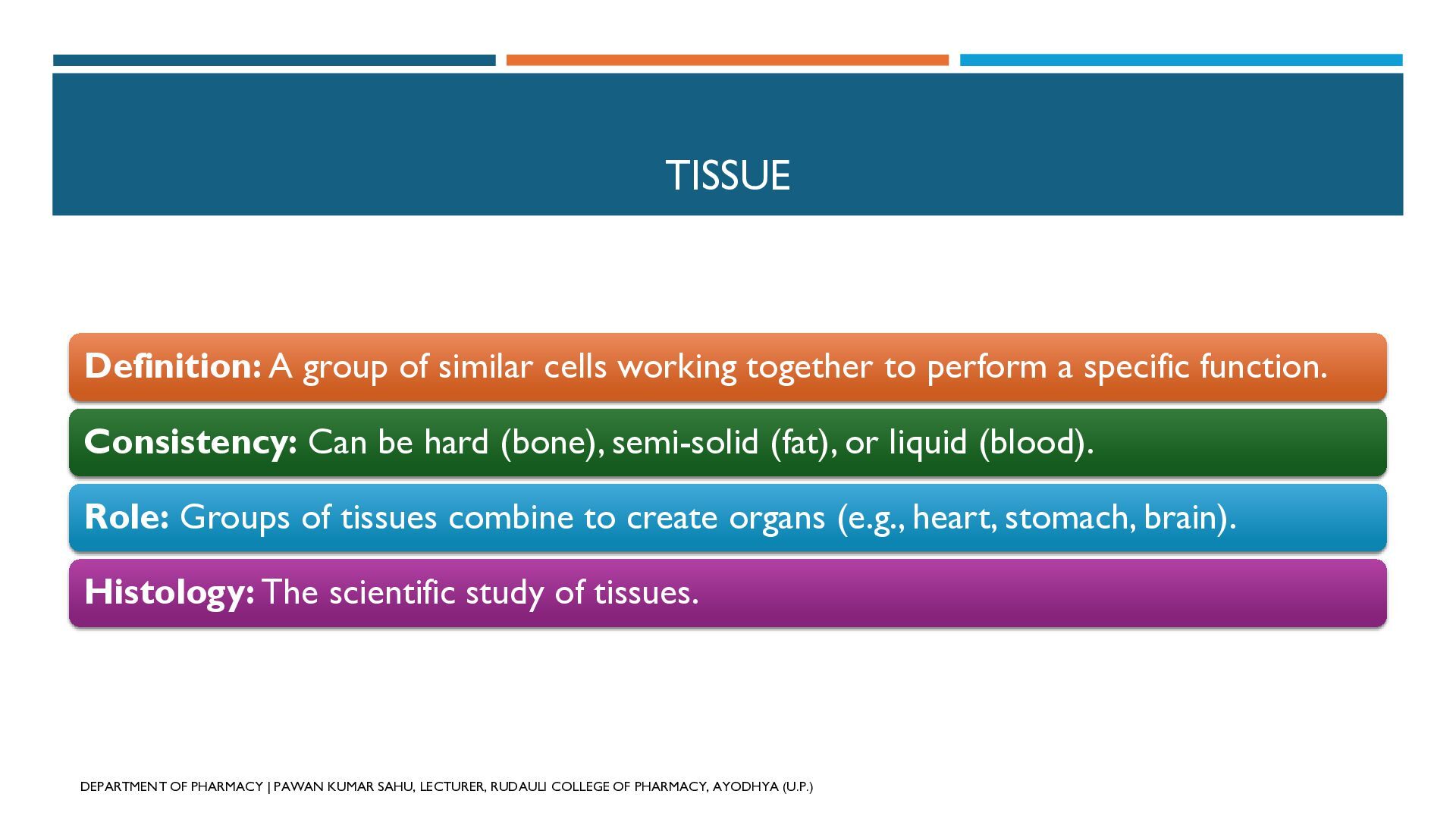
  1. TISSUE Definition: A group of similar cells working together to

    perform a specific function. Consistency: Can be hard (bone), semi-solid (fat), or liquid (blood). Role: Groups of tissues combine to create organs (e.g., heart, stomach, brain). Histology: The scientific study of tissues. DEPARTMENT OF PHARMACY | PAWAN KUMAR SAHU, LECTURER, RUDAULI COLLEGE OF PHARMACY, AYODHYA (U.P.)
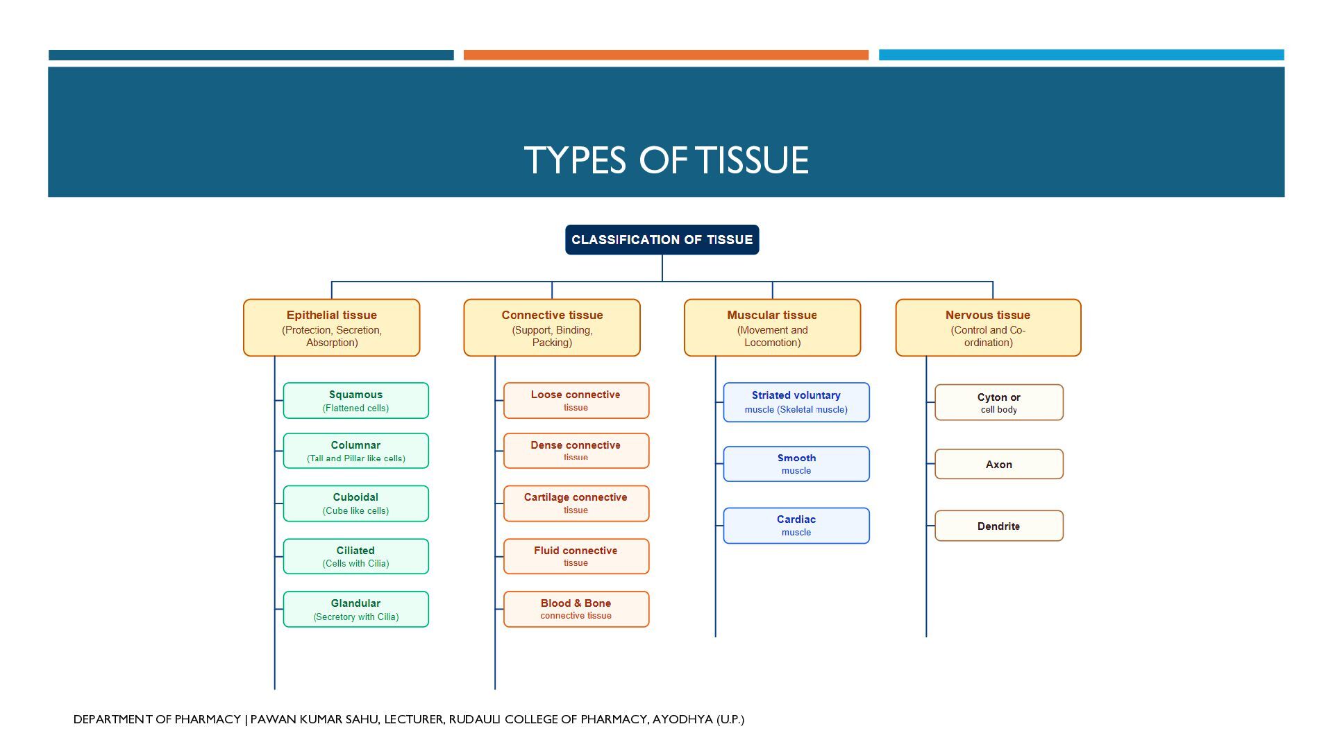
  2. TYPES OF TISSUE DEPARTMENT OF PHARMACY | PAWAN KUMAR SAHU,

    LECTURER, RUDAULI COLLEGE OF PHARMACY, AYODHYA (U.P.)
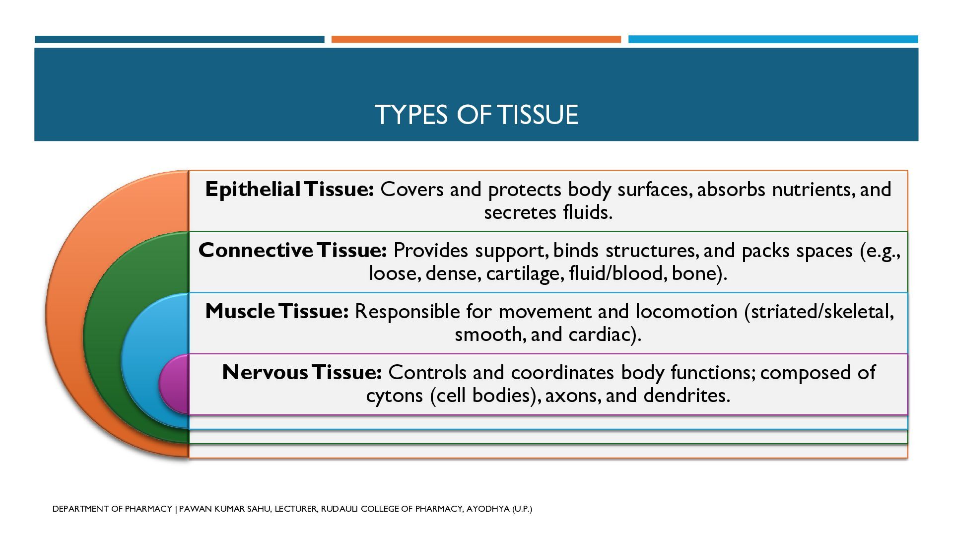
  3. TYPES OF TISSUE Epithelial Tissue: Covers and protects body surfaces,

    absorbs nutrients, and secretes fluids. Connective Tissue: Provides support, binds structures, and packs spaces (e.g., loose, dense, cartilage, fluid/blood, bone). Muscle Tissue: Responsible for movement and locomotion (striated/skeletal, smooth, and cardiac). Nervous Tissue: Controls and coordinates body functions; composed of cytons (cell bodies), axons, and dendrites. DEPARTMENT OF PHARMACY | PAWAN KUMAR SAHU, LECTURER, RUDAULI COLLEGE OF PHARMACY, AYODHYA (U.P.)
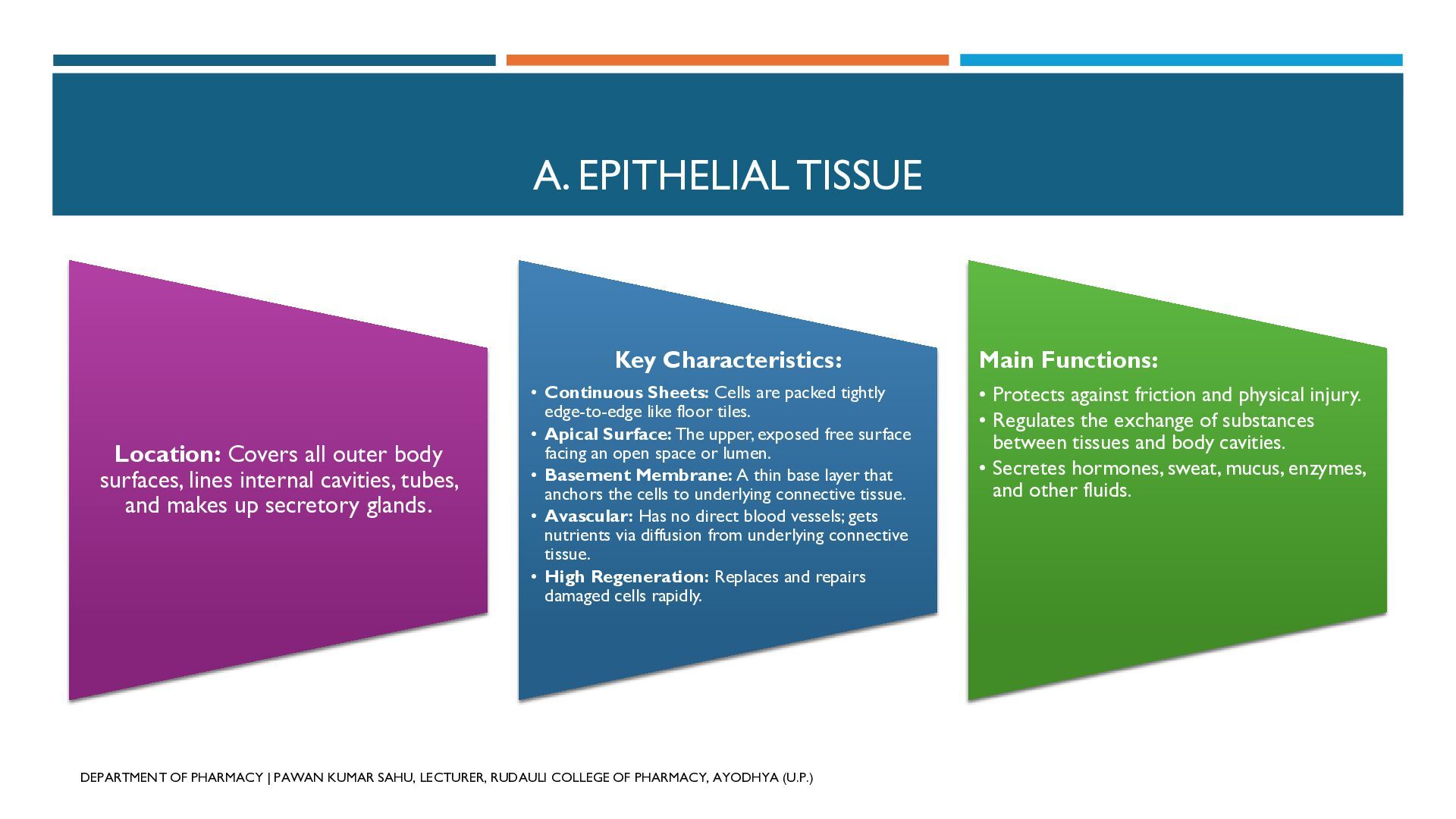
  4. A. EPITHELIAL TISSUE Key Characteristics: Location: Covers all outer body

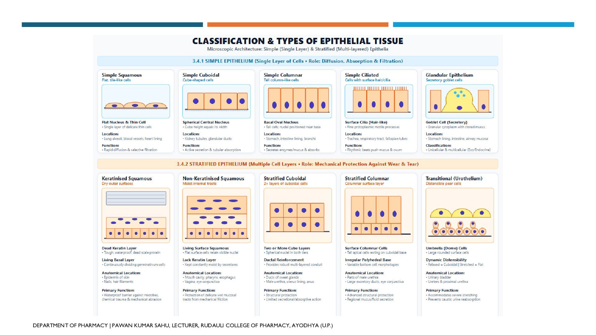
    surfaces, lines internal cavities, tubes, and makes up secretory glands. • Continuous Sheets: Cells are packed tightly edge-to-edge like floor tiles. • Apical Surface: The upper, exposed free surface facing an open space or lumen. • Basement Membrane: A thin base layer that anchors the cells to underlying connective tissue. • Avascular: Has no direct blood vessels; gets nutrients via diffusion from underlying connective tissue. • High Regeneration: Replaces and repairs damaged cells rapidly. DEPARTMENT OF PHARMACY | PAWAN KUMAR SAHU, LECTURER, RUDAULI COLLEGE OF PHARMACY, AYODHYA (U.P.) Main Functions: • Protects against friction and physical injury. • Regulates the exchange of substances between tissues and body cavities. • Secretes hormones, sweat, mucus, enzymes, and other fluids.
  5. TYPES OF EPITHELIAL TISSUE DEPARTMENT OF PHARMACY | PAWAN KUMAR

    SAHU, LECTURER, RUDAULI COLLEGE OF PHARMACY, AYODHYA (U.P.)
  6. 1. SIMPLE EPITHELIAL TISSUES a) Simple Squamous Epithelium • Locations:

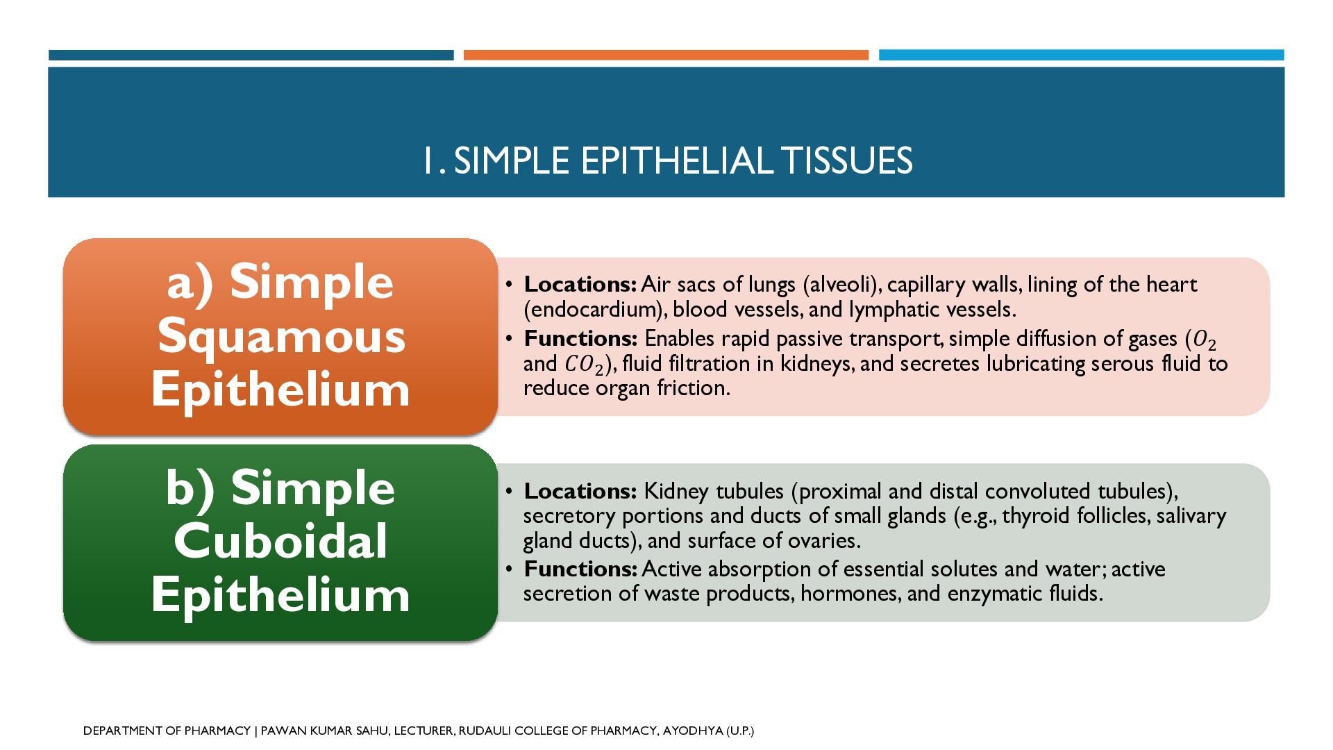
    Air sacs of lungs (alveoli), capillary walls, lining of the heart (endocardium), blood vessels, and lymphatic vessels. • Functions: Enables rapid passive transport, simple diffusion of gases (𝑂2 and 𝐶𝑂2 ), fluid filtration in kidneys, and secretes lubricating serous fluid to reduce organ friction. b) Simple Cuboidal Epithelium • Locations: Kidney tubules (proximal and distal convoluted tubules), secretory portions and ducts of small glands (e.g., thyroid follicles, salivary gland ducts), and surface of ovaries. • Functions: Active absorption of essential solutes and water; active secretion of waste products, hormones, and enzymatic fluids. cap O sub 2 cap C cap O sub 2 DEPARTMENT OF PHARMACY | PAWAN KUMAR SAHU, LECTURER, RUDAULI COLLEGE OF PHARMACY, AYODHYA (U.P.)
  7. 1. SIMPLE EPITHELIAL TISSUES c) Simple Columnar Epithelium • Locations:

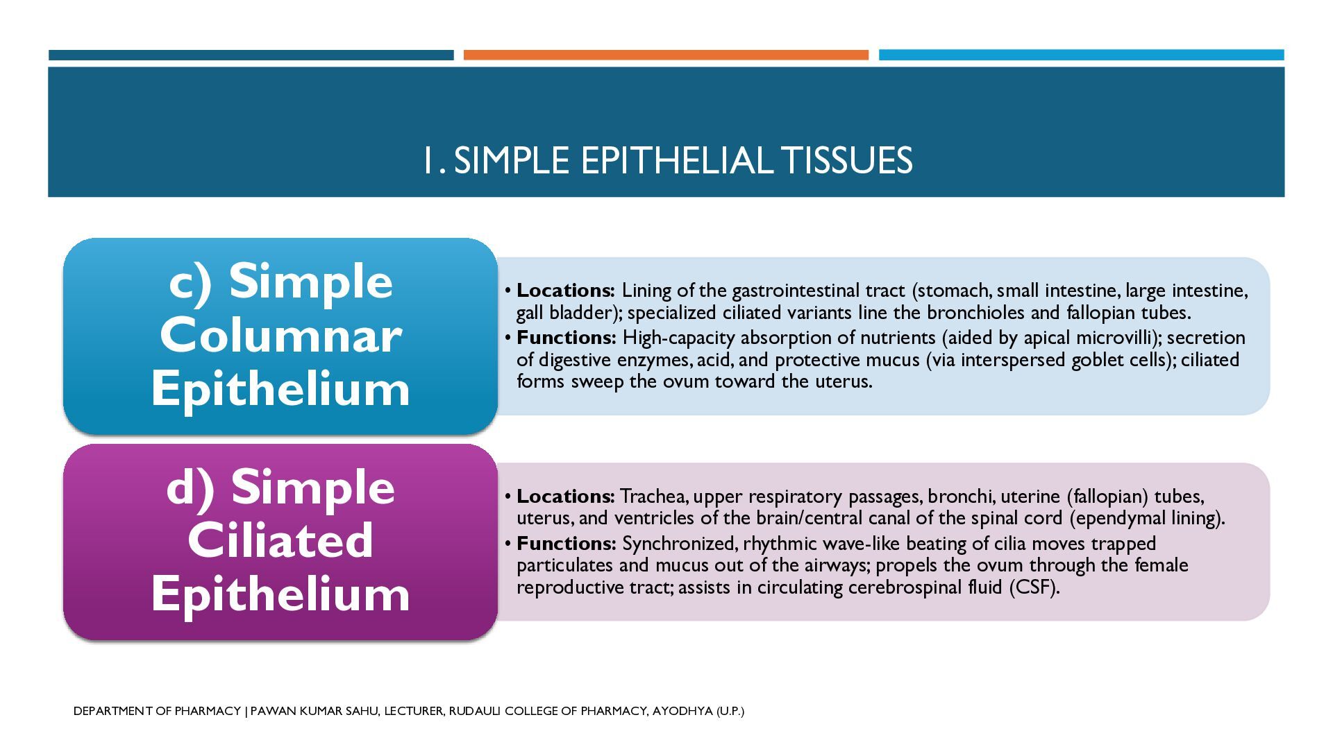
    Lining of the gastrointestinal tract (stomach, small intestine, large intestine, gall bladder); specialized ciliated variants line the bronchioles and fallopian tubes. • Functions: High-capacity absorption of nutrients (aided by apical microvilli); secretion of digestive enzymes, acid, and protective mucus (via interspersed goblet cells); ciliated forms sweep the ovum toward the uterus. d) Simple Ciliated Epithelium • Locations: Trachea, upper respiratory passages, bronchi, uterine (fallopian) tubes, uterus, and ventricles of the brain/central canal of the spinal cord (ependymal lining). • Functions: Synchronized, rhythmic wave-like beating of cilia moves trapped particulates and mucus out of the airways; propels the ovum through the female reproductive tract; assists in circulating cerebrospinal fluid (CSF). DEPARTMENT OF PHARMACY | PAWAN KUMAR SAHU, LECTURER, RUDAULI COLLEGE OF PHARMACY, AYODHYA (U.P.)
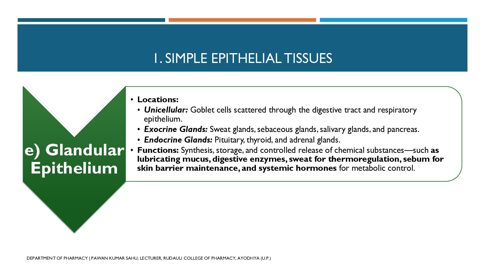
  8. 1. SIMPLE EPITHELIAL TISSUES e) Glandular Epithelium • Locations: •

    Unicellular: Goblet cells scattered through the digestive tract and respiratory epithelium. • Exocrine Glands: Sweat glands, sebaceous glands, salivary glands, and pancreas. • Endocrine Glands: Pituitary, thyroid, and adrenal glands. • Functions: Synthesis, storage, and controlled release of chemical substances—such as lubricating mucus, digestive enzymes, sweat for thermoregulation, sebum for skin barrier maintenance, and systemic hormones for metabolic control. DEPARTMENT OF PHARMACY | PAWAN KUMAR SAHU, LECTURER, RUDAULI COLLEGE OF PHARMACY, AYODHYA (U.P.)
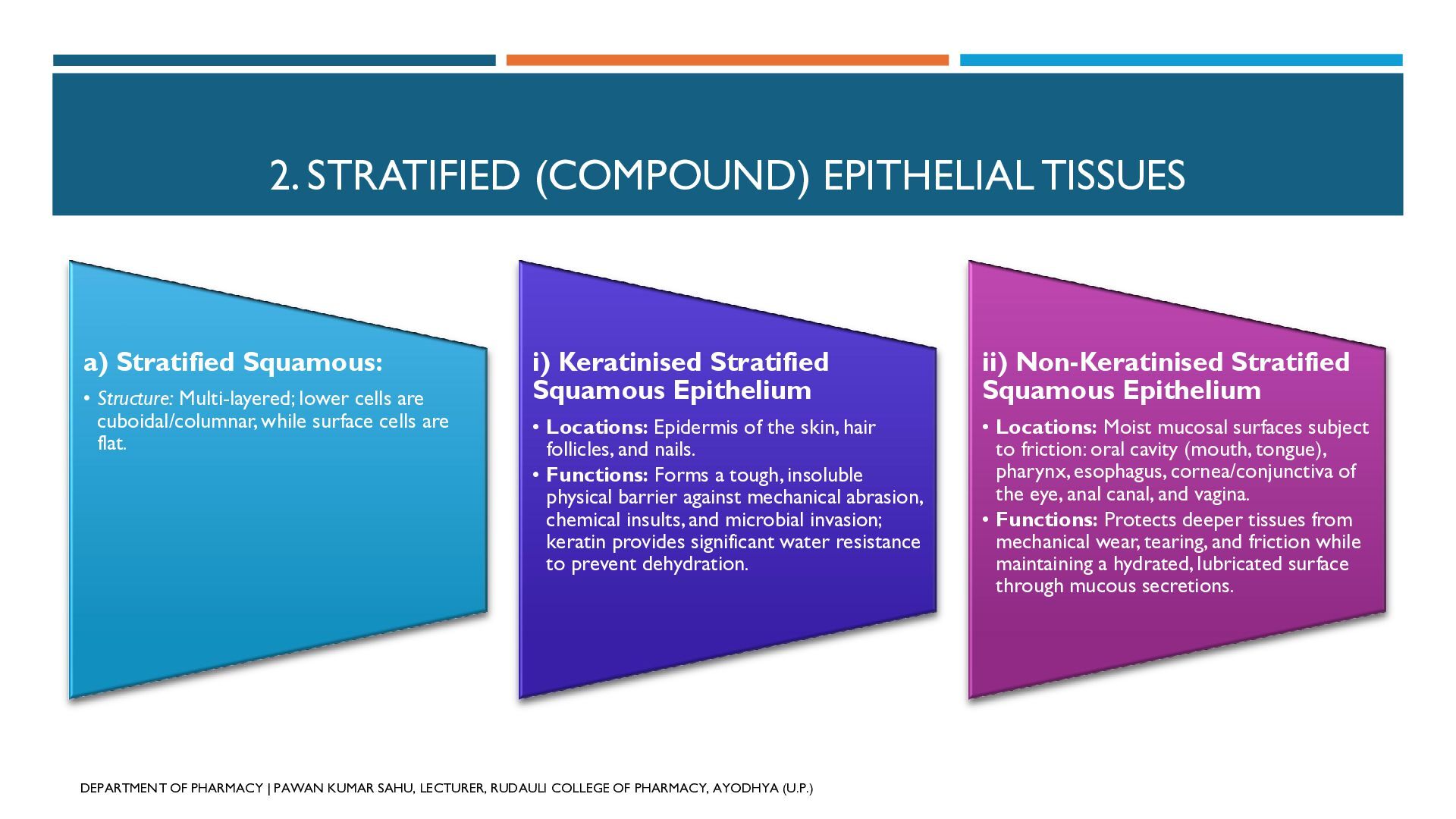
  9. 2. STRATIFIED (COMPOUND) EPITHELIAL TISSUES a) Stratified Squamous: • Structure:

    Multi-layered; lower cells are cuboidal/columnar, while surface cells are flat. i) Keratinised Stratified Squamous Epithelium ii) Non-Keratinised Stratified Squamous Epithelium • Locations: Epidermis of the skin, hair follicles, and nails. • Functions: Forms a tough, insoluble physical barrier against mechanical abrasion, chemical insults, and microbial invasion; keratin provides significant water resistance to prevent dehydration. • Locations: Moist mucosal surfaces subject to friction: oral cavity (mouth, tongue), pharynx, esophagus, cornea/conjunctiva of the eye, anal canal, and vagina. • Functions: Protects deeper tissues from mechanical wear, tearing, and friction while maintaining a hydrated, lubricated surface through mucous secretions. DEPARTMENT OF PHARMACY | PAWAN KUMAR SAHU, LECTURER, RUDAULI COLLEGE OF PHARMACY, AYODHYA (U.P.)
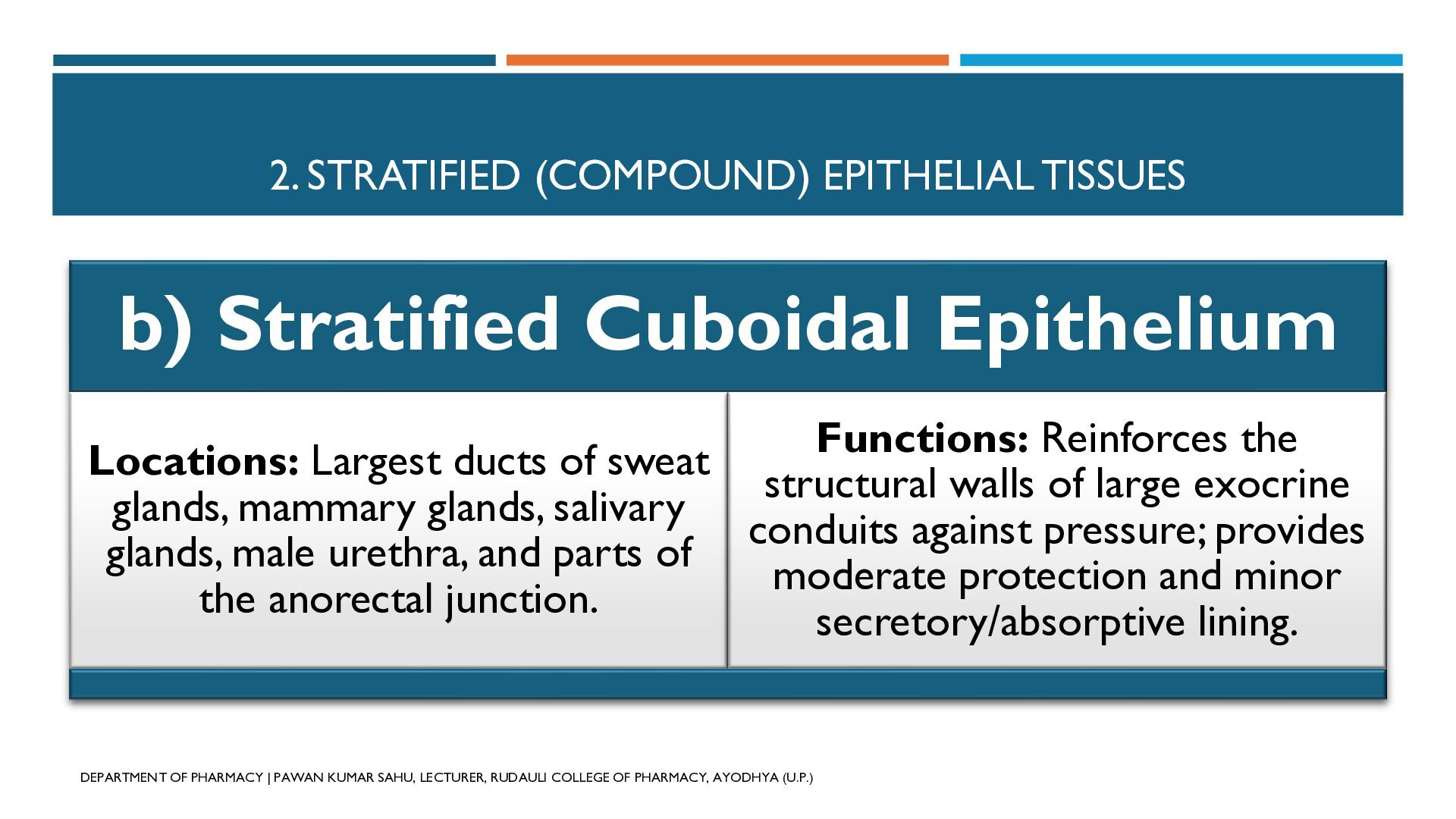
  10. 2. STRATIFIED (COMPOUND) EPITHELIAL TISSUES b) Stratified Cuboidal Epithelium Functions:

    Reinforces the Locations: Largest ducts of sweat structural walls of large exocrine glands, mammary glands, salivary conduits against pressure; provides glands, male urethra, and parts of moderate protection and minor the anorectal junction. secretory/absorptive lining. DEPARTMENT OF PHARMACY | PAWAN KUMAR SAHU, LECTURER, RUDAULI COLLEGE OF PHARMACY, AYODHYA (U.P.)
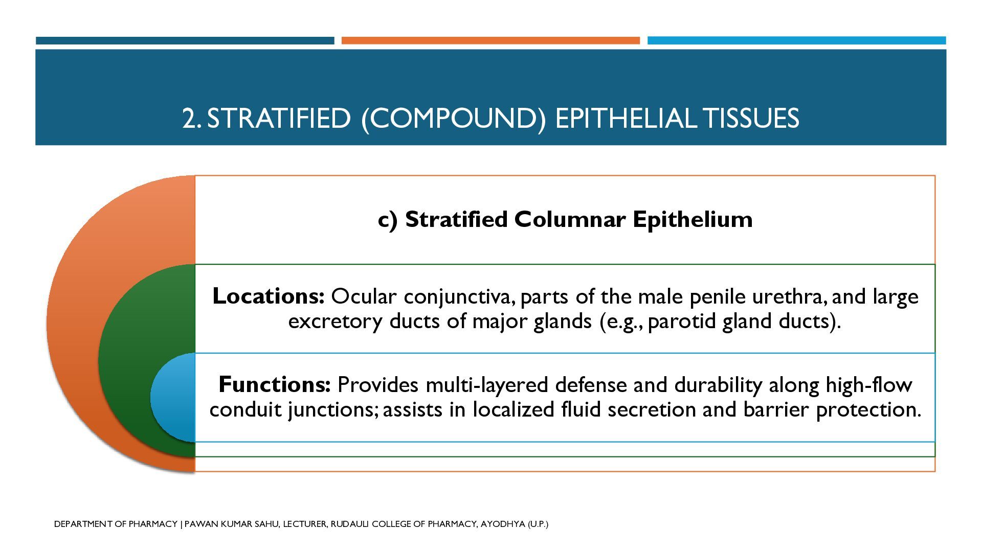
  11. 2. STRATIFIED (COMPOUND) EPITHELIAL TISSUES c) Stratified Columnar Epithelium Locations:

    Ocular conjunctiva, parts of the male penile urethra, and large excretory ducts of major glands (e.g., parotid gland ducts). Functions: Provides multi-layered defense and durability along high-flow conduit junctions; assists in localized fluid secretion and barrier protection. DEPARTMENT OF PHARMACY | PAWAN KUMAR SAHU, LECTURER, RUDAULI COLLEGE OF PHARMACY, AYODHYA (U.P.)
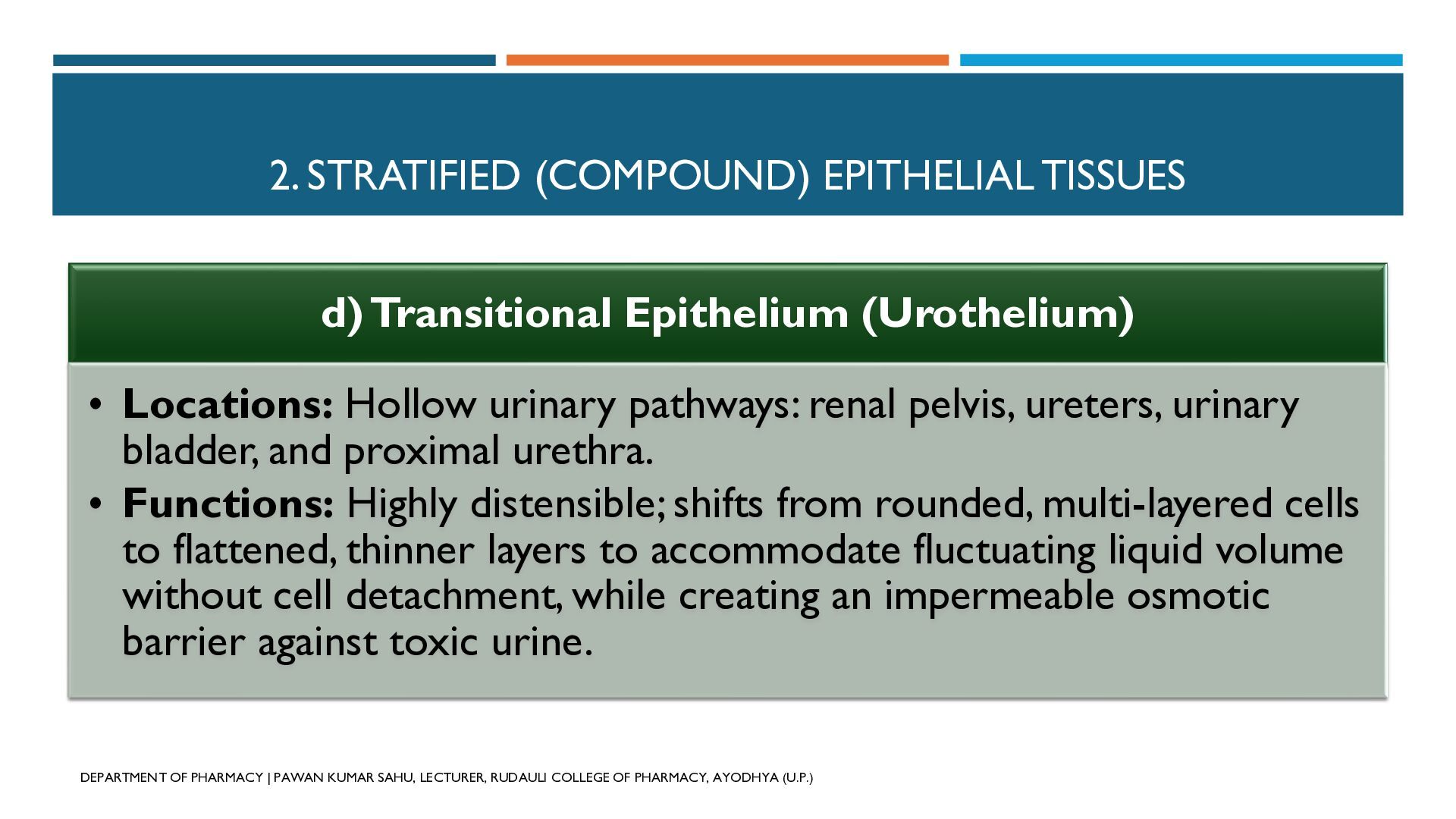
  12. 2. STRATIFIED (COMPOUND) EPITHELIAL TISSUES d) Transitional Epithelium (Urothelium) •

    Locations: Hollow urinary pathways: renal pelvis, ureters, urinary bladder, and proximal urethra. • Functions: Highly distensible; shifts from rounded, multi-layered cells to flattened, thinner layers to accommodate fluctuating liquid volume without cell detachment, while creating an impermeable osmotic barrier against toxic urine. DEPARTMENT OF PHARMACY | PAWAN KUMAR SAHU, LECTURER, RUDAULI COLLEGE OF PHARMACY, AYODHYA (U.P.)
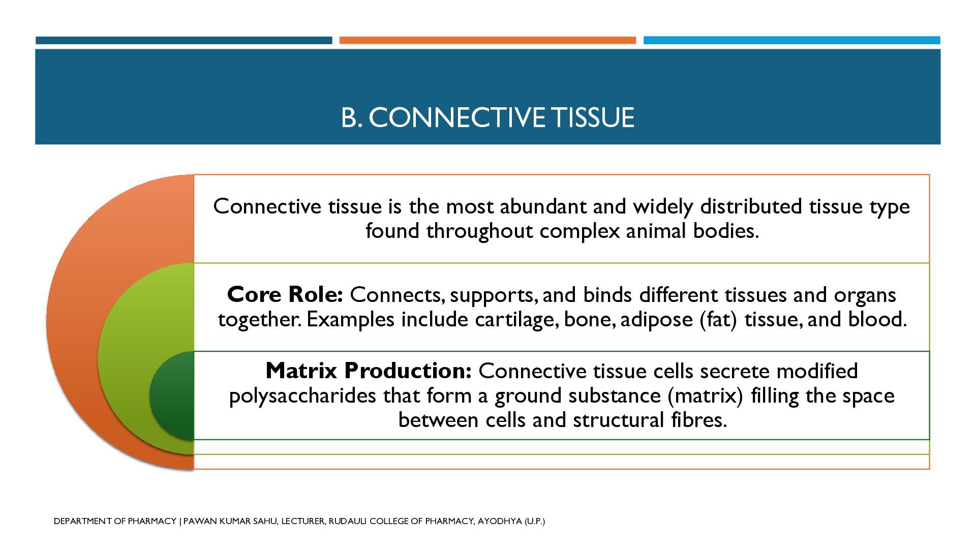
  13. B. CONNECTIVE TISSUE Connective tissue is the most abundant and

    widely distributed tissue type found throughout complex animal bodies. Core Role: Connects, supports, and binds different tissues and organs together. Examples include cartilage, bone, adipose (fat) tissue, and blood. Matrix Production: Connective tissue cells secrete modified polysaccharides that form a ground substance (matrix) filling the space between cells and structural fibres. DEPARTMENT OF PHARMACY | PAWAN KUMAR SAHU, LECTURER, RUDAULI COLLEGE OF PHARMACY, AYODHYA (U.P.)
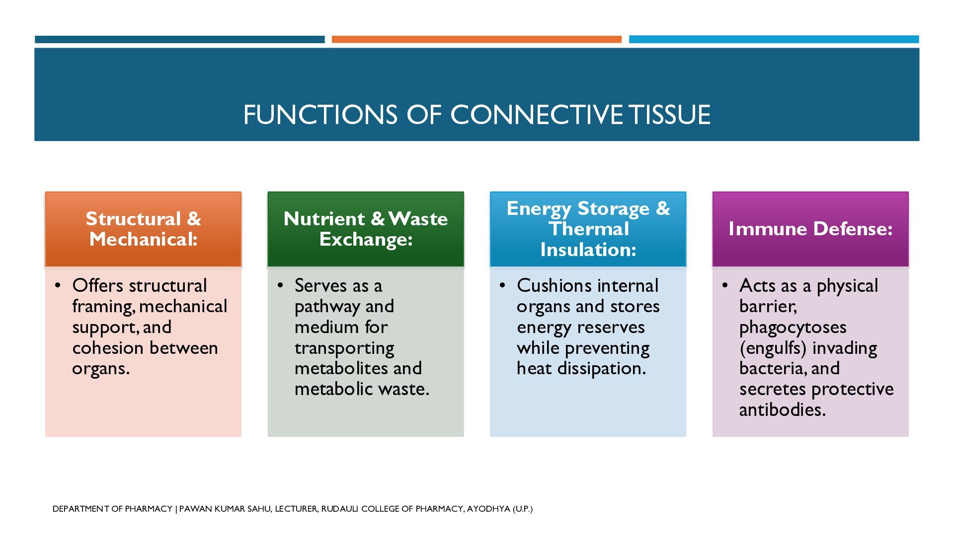
  14. FUNCTIONS OF CONNECTIVE TISSUE Structural & Mechanical: Nutrient & Waste

    Exchange: • Offers structural framing, mechanical support, and cohesion between organs. • Serves as a pathway and medium for transporting metabolites and metabolic waste. Energy Storage & Thermal Insulation: • Cushions internal organs and stores energy reserves while preventing heat dissipation. DEPARTMENT OF PHARMACY | PAWAN KUMAR SAHU, LECTURER, RUDAULI COLLEGE OF PHARMACY, AYODHYA (U.P.) Immune Defense: • Acts as a physical barrier, phagocytoses (engulfs) invading bacteria, and secretes protective antibodies.
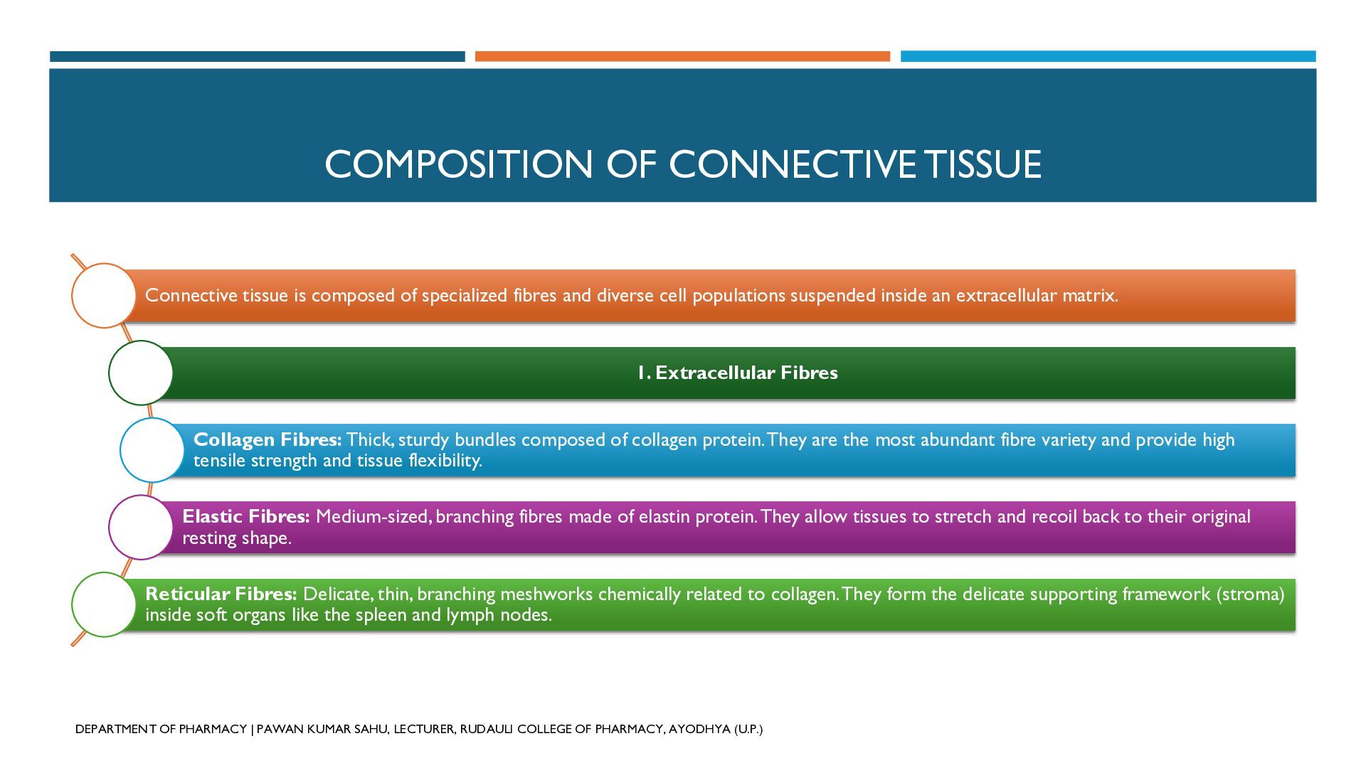
  15. COMPOSITION OF CONNECTIVE TISSUE Connective tissue is composed of specialized

    fibres and diverse cell populations suspended inside an extracellular matrix. 1. Extracellular Fibres Collagen Fibres: Thick, sturdy bundles composed of collagen protein.They are the most abundant fibre variety and provide high tensile strength and tissue flexibility. Elastic Fibres: Medium-sized, branching fibres made of elastin protein. They allow tissues to stretch and recoil back to their original resting shape. Reticular Fibres: Delicate, thin, branching meshworks chemically related to collagen.They form the delicate supporting framework (stroma) inside soft organs like the spleen and lymph nodes. DEPARTMENT OF PHARMACY | PAWAN KUMAR SAHU, LECTURER, RUDAULI COLLEGE OF PHARMACY, AYODHYA (U.P.)
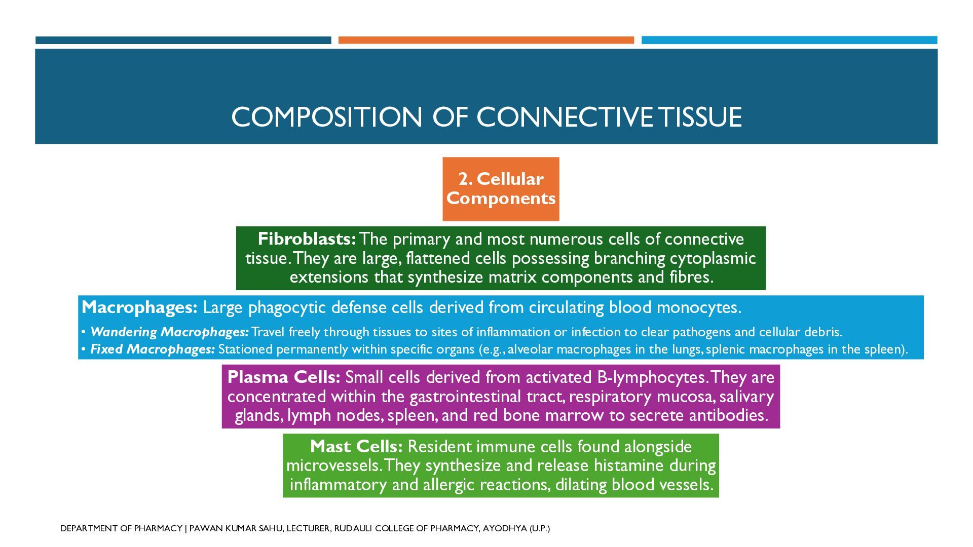
  16. COMPOSITION OF CONNECTIVE TISSUE 2. Cellular Components Fibroblasts: The primary

    and most numerous cells of connective tissue. They are large, flattened cells possessing branching cytoplasmic extensions that synthesize matrix components and fibres. Macrophages: Large phagocytic defense cells derived from circulating blood monocytes. • Wandering Macrophages: Travel freely through tissues to sites of inflammation or infection to clear pathogens and cellular debris. • Fixed Macrophages: Stationed permanently within specific organs (e.g., alveolar macrophages in the lungs, splenic macrophages in the spleen). Plasma Cells: Small cells derived from activated B-lymphocytes. They are concentrated within the gastrointestinal tract, respiratory mucosa, salivary glands, lymph nodes, spleen, and red bone marrow to secrete antibodies. Mast Cells: Resident immune cells found alongside microvessels. They synthesize and release histamine during inflammatory and allergic reactions, dilating blood vessels. DEPARTMENT OF PHARMACY | PAWAN KUMAR SAHU, LECTURER, RUDAULI COLLEGE OF PHARMACY, AYODHYA (U.P.)
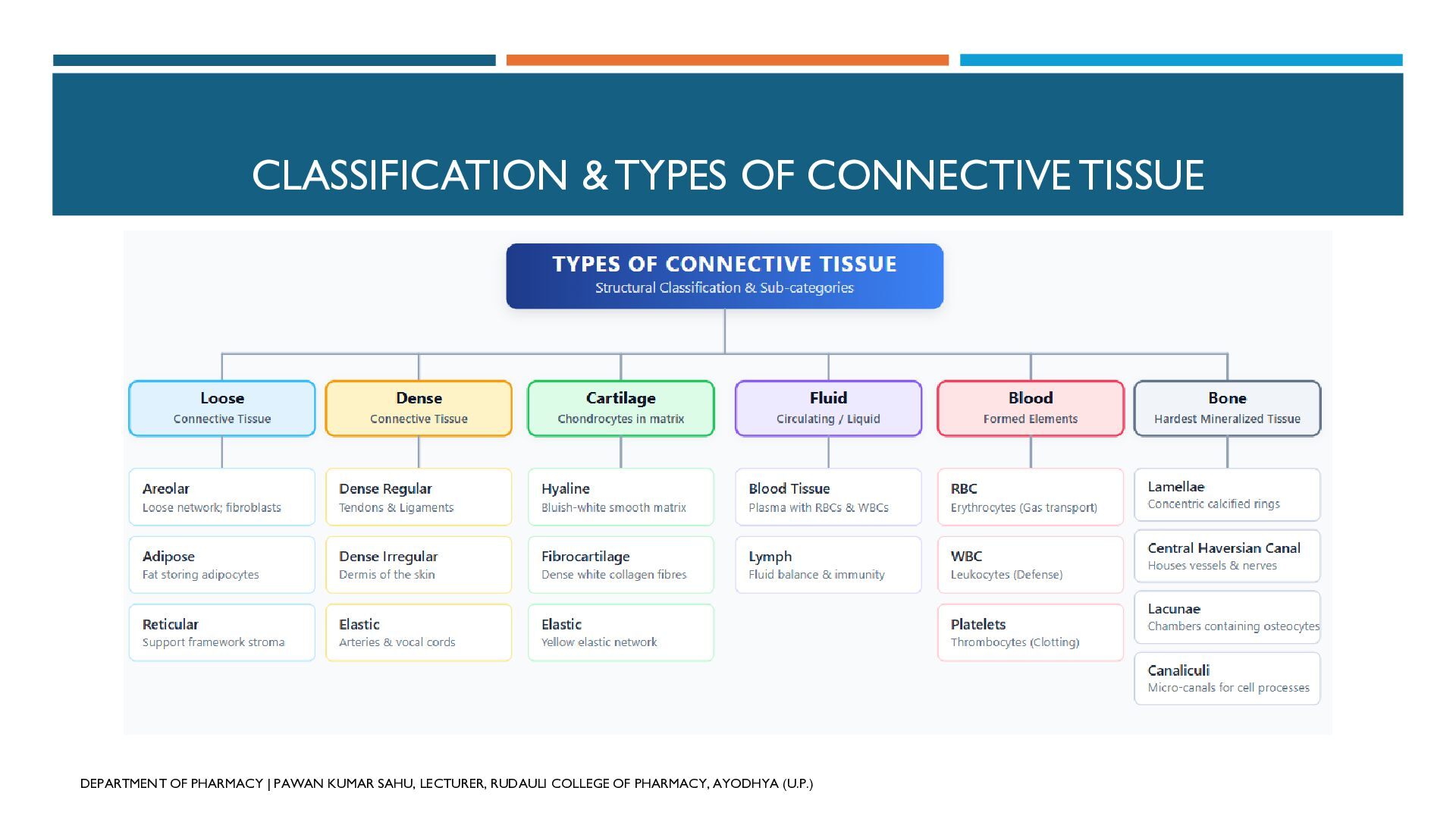
  17. CLASSIFICATION & TYPES OF CONNECTIVE TISSUE DEPARTMENT OF PHARMACY |

    PAWAN KUMAR SAHU, LECTURER, RUDAULI COLLEGE OF PHARMACY, AYODHYA (U.P.)
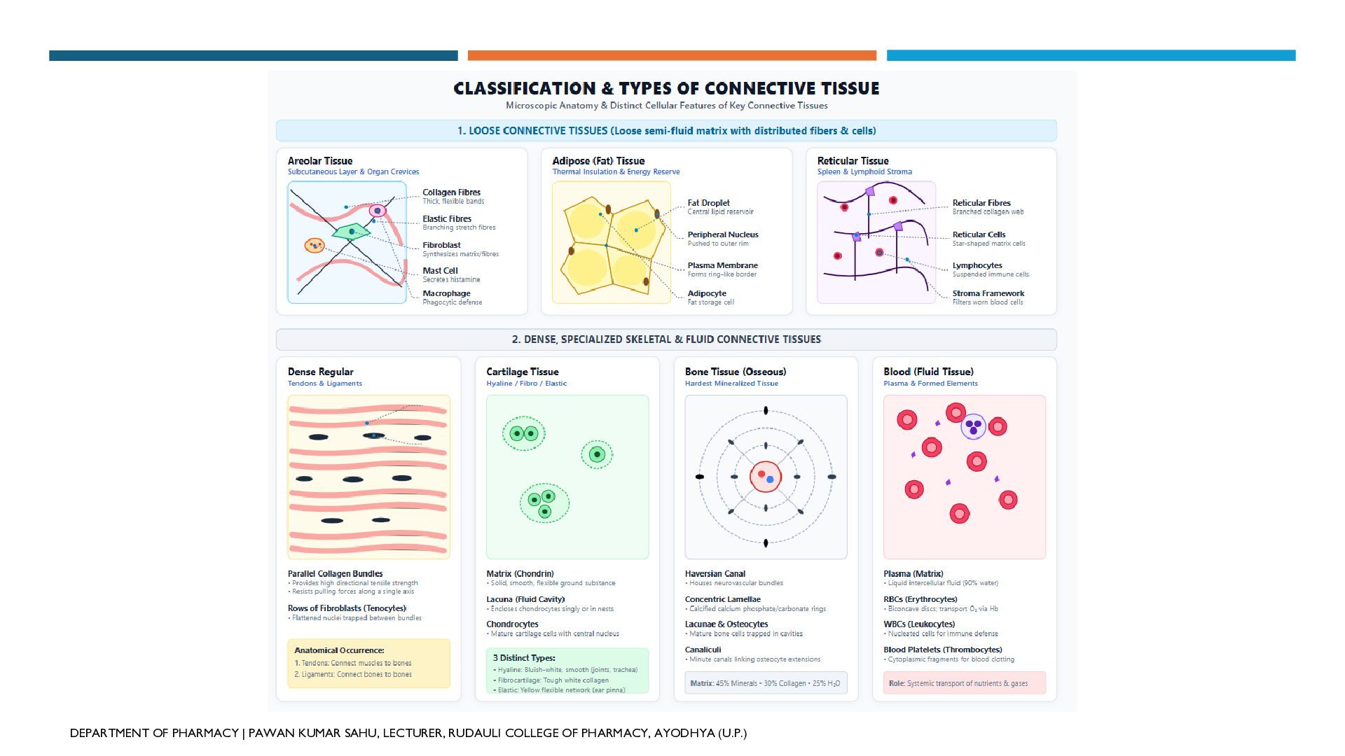
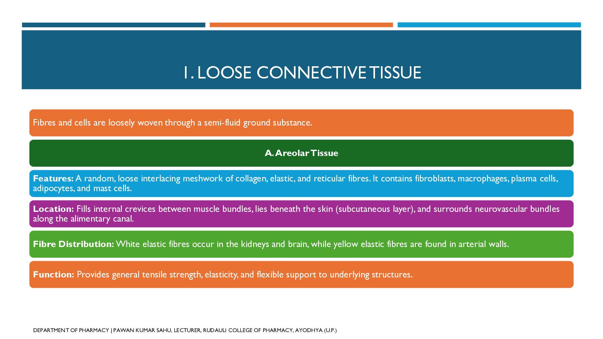
  18. 1. LOOSE CONNECTIVE TISSUE Fibres and cells are loosely woven

    through a semi-fluid ground substance. A. Areolar Tissue Features: A random, loose interlacing meshwork of collagen, elastic, and reticular fibres. It contains fibroblasts, macrophages, plasma cells, adipocytes, and mast cells. Location: Fills internal crevices between muscle bundles, lies beneath the skin (subcutaneous layer), and surrounds neurovascular bundles along the alimentary canal. Fibre Distribution: White elastic fibres occur in the kidneys and brain, while yellow elastic fibres are found in arterial walls. Function: Provides general tensile strength, elasticity, and flexible support to underlying structures. DEPARTMENT OF PHARMACY | PAWAN KUMAR SAHU, LECTURER, RUDAULI COLLEGE OF PHARMACY, AYODHYA (U.P.)
  19. 1. LOOSE CONNECTIVE TISSUE B. Adipose Tissue Features: Composed of

    specialized fat-storing cells called adipocytes. Each adipocyte contains a large central lipid droplet that pushes the cytoplasm and nucleus to the periphery. Location: Deep subcutaneous tissue beneath the skin, around vulnerable internal organs (heart, kidneys), and inside yellow bone marrow. Function: Serves as a major calorie reservoir, prevents loss of body heat (thermal insulation), absorbs mechanical shocks, and shapes body contours. DEPARTMENT OF PHARMACY | PAWAN KUMAR SAHU, LECTURER, RUDAULI COLLEGE OF PHARMACY, AYODHYA (U.P.)
  20. 1. LOOSE CONNECTIVE TISSUE C. Reticular Connective Tissue Features: A

    delicate network of fine reticular fibres populated by reticular cells. Location: Supporting stroma of the liver, spleen, lymph nodes, and red bone marrow; also surrounds muscle fibres and capillaries. DEPARTMENT OF PHARMACY | PAWAN KUMAR SAHU, LECTURER, RUDAULI COLLEGE OF PHARMACY, AYODHYA (U.P.) Function: Forms a structural filtration mesh inside lymphoid organs that traps wornout red blood cells (in the spleen) and filters foreign microbes (in lymph nodes).
  21. 2. DENSE CONNECTIVE TISSUE Dominated by tightly packed, dense protein

    fibres with fewer cells and reduced matrix volume. A. Dense Regular Connective Tissue Features: Parallel arrays and bundles of collagen fibres arranged neatly in straight, alternating rows. DEPARTMENT OF PHARMACY | PAWAN KUMAR SAHU, LECTURER, RUDAULI COLLEGE OF PHARMACY, AYODHYA (U.P.) Location: • Tendons: Connect skeletal muscles firmly to bones. • Ligaments: Connect bones to other bones across joints. Function: Delivers high directional tensile strength and resists pulling forces along a single axis.
  22. 2. DENSE CONNECTIVE TISSUE B. Dense Irregular Connective Tissue Features:

    Dense interwoven bundles of collagen fibres oriented randomly in multiple directions, interspersed with fibroblasts. Location: Found prominently within the deeper dermis of the skin. Function: Resists multidirectional mechanical tension and prevents tearing. C. Dense Elastic Connective Tissue Features: Branching networks of elastic fibres accompanied by scattered fibroblasts; distinctly yellowish in color. Location: Walls of large elastic arteries, bronchial tubes, lung parenchyma, trachea, and vocal cords. Function: Permits substantial expansion and elastic recoil in organs subjected to cyclical pressure changes. DEPARTMENT OF PHARMACY | PAWAN KUMAR SAHU, LECTURER, RUDAULI COLLEGE OF PHARMACY, AYODHYA (U.P.)
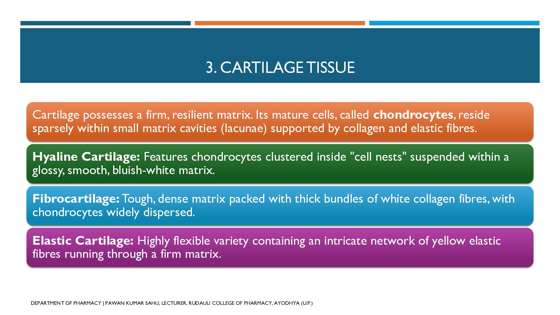
  23. 3. CARTILAGE TISSUE Cartilage possesses a firm, resilient matrix. Its

    mature cells, called chondrocytes, reside sparsely within small matrix cavities (lacunae) supported by collagen and elastic fibres. Hyaline Cartilage: Features chondrocytes clustered inside "cell nests" suspended within a glossy, smooth, bluish-white matrix. Fibrocartilage: Tough, dense matrix packed with thick bundles of white collagen fibres, with chondrocytes widely dispersed. Elastic Cartilage: Highly flexible variety containing an intricate network of yellow elastic fibres running through a firm matrix. DEPARTMENT OF PHARMACY | PAWAN KUMAR SAHU, LECTURER, RUDAULI COLLEGE OF PHARMACY, AYODHYA (U.P.)
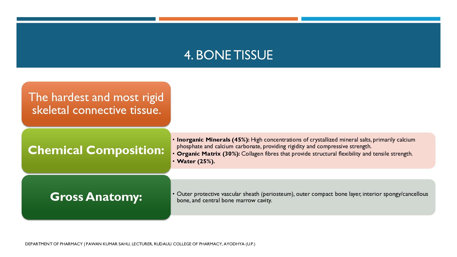
  24. 4. BONE TISSUE The hardest and most rigid skeletal connective

    tissue. Chemical Composition: Gross Anatomy: • Inorganic Minerals (45%): High concentrations of crystallized mineral salts, primarily calcium phosphate and calcium carbonate, providing rigidity and compressive strength. • Organic Matrix (30%): Collagen fibres that provide structural flexibility and tensile strength. • Water (25%). • Outer protective vascular sheath (periosteum), outer compact bone layer, interior spongy/cancellous bone, and central bone marrow cavity. DEPARTMENT OF PHARMACY | PAWAN KUMAR SAHU, LECTURER, RUDAULI COLLEGE OF PHARMACY, AYODHYA (U.P.)
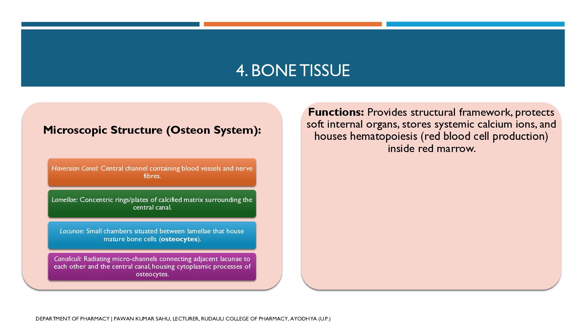
  25. 4. BONE TISSUE Microscopic Structure (Osteon System): Functions: Provides structural

    framework, protects soft internal organs, stores systemic calcium ions, and houses hematopoiesis (red blood cell production) inside red marrow. Haversian Canal: Central channel containing blood vessels and nerve fibres. Lamellae: Concentric rings/plates of calcified matrix surrounding the central canal. Lacunae: Small chambers situated between lamellae that house mature bone cells (osteocytes). Canaliculi: Radiating micro-channels connecting adjacent lacunae to each other and the central canal, housing cytoplasmic processes of osteocytes. DEPARTMENT OF PHARMACY | PAWAN KUMAR SAHU, LECTURER, RUDAULI COLLEGE OF PHARMACY, AYODHYA (U.P.)
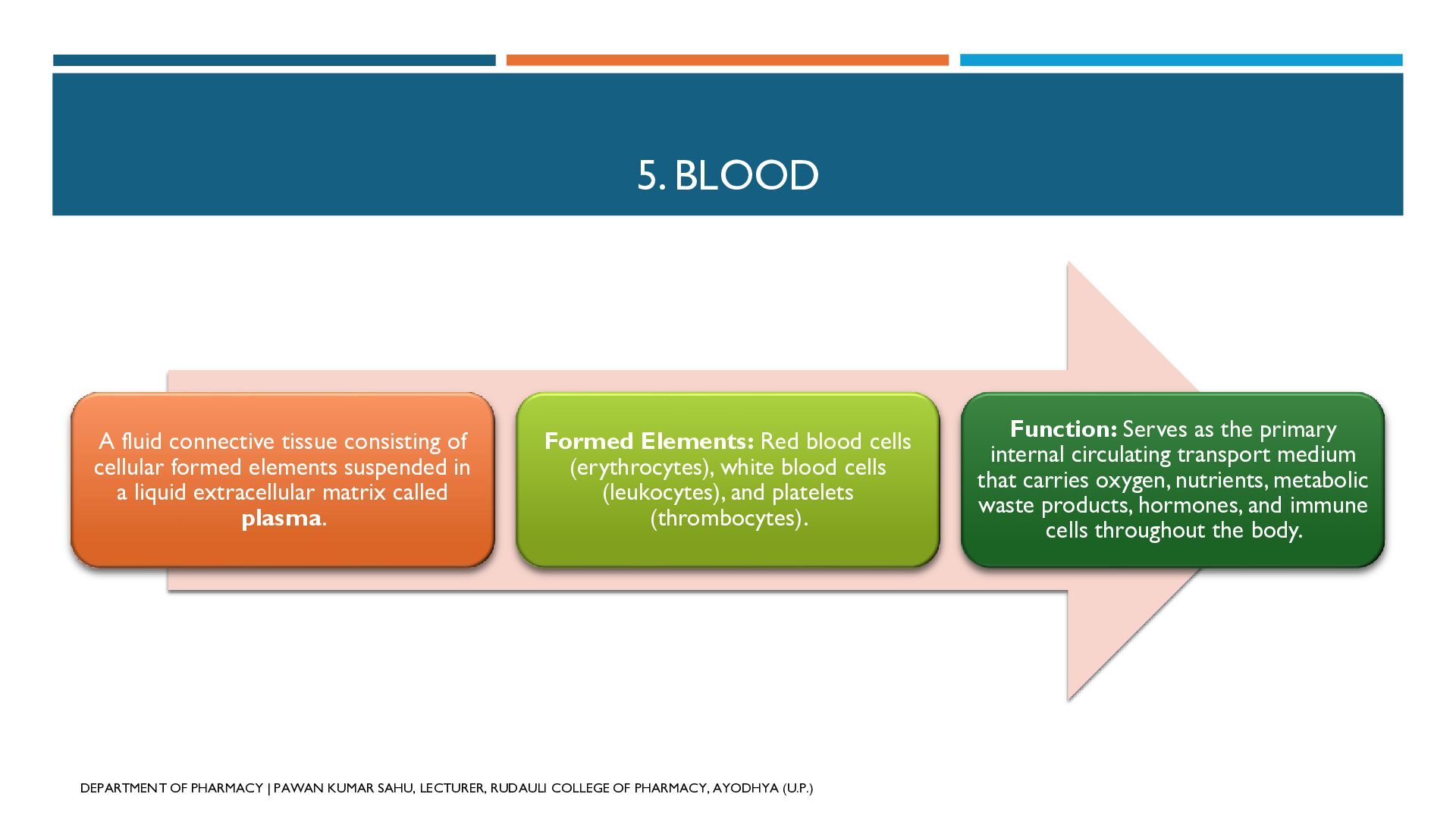
  26. 5. BLOOD A fluid connective tissue consisting of cellular formed

    elements suspended in a liquid extracellular matrix called plasma. Formed Elements: Red blood cells (erythrocytes), white blood cells (leukocytes), and platelets (thrombocytes). DEPARTMENT OF PHARMACY | PAWAN KUMAR SAHU, LECTURER, RUDAULI COLLEGE OF PHARMACY, AYODHYA (U.P.) Function: Serves as the primary internal circulating transport medium that carries oxygen, nutrients, metabolic waste products, hormones, and immune cells throughout the body.
  27. C. MUSCLE TISSUE Composed of elongated, contractile muscle cells called

    muscle fibres arranged in parallel arrays. Each fibre is packed internally with smaller contractile units called myofibrils. When stimulated, these fibres contract and relax in an organized manner. Functions: Enables active body movement and locomotion, maintains posture, stabilizes joint positions, and shields deeper viscera from mechanical stress. DEPARTMENT OF PHARMACY | PAWAN KUMAR SAHU, LECTURER, RUDAULI COLLEGE OF PHARMACY, AYODHYA (U.P.)
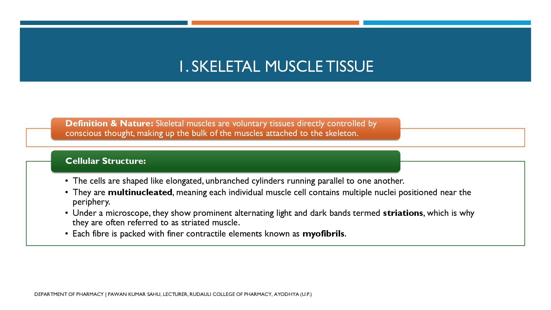
  28. 1. SKELETAL MUSCLE TISSUE Definition & Nature: Skeletal muscles are

    voluntary tissues directly controlled by conscious thought, making up the bulk of the muscles attached to the skeleton. Cellular Structure: • The cells are shaped like elongated, unbranched cylinders running parallel to one another. • They are multinucleated, meaning each individual muscle cell contains multiple nuclei positioned near the periphery. • Under a microscope, they show prominent alternating light and dark bands termed striations, which is why they are often referred to as striated muscle. • Each fibre is packed with finer contractile elements known as myofibrils. DEPARTMENT OF PHARMACY | PAWAN KUMAR SAHU, LECTURER, RUDAULI COLLEGE OF PHARMACY, AYODHYA (U.P.)
  29. 1. SKELETAL MUSCLE TISSUE Location: They are attached securely to

    the skeletal bones across joints by strong bands of fibrous connective tissue called tendons. Functions: • Drives active locomotion (walking, running) and voluntary limb movements. • Controls facial expressions and body gestures. • Stabilizes joints, maintains body posture, and shields underlying visceral structures from mechanical impacts. DEPARTMENT OF PHARMACY | PAWAN KUMAR SAHU, LECTURER, RUDAULI COLLEGE OF PHARMACY, AYODHYA (U.P.)
  30. 2. CARDIAC MUSCLE TISSUE Definition & Nature: Cardiac muscle is

    a specialized, involuntary striated muscle engineered for uninterrupted rhythmic performance without undergoing fatigue. Cellular Structure: • The fibres are cylindrical and uniquely branched, forming a connected web-like structure. • Unlike skeletal fibres, cardiac cells typically feature a single centrally located nucleus (uninucleate). • They show faint striations and are joined end-to-end through specialized junctional membranes called intercalated discs. These discs permit electrical impulses to pass rapidly from one cell to another so that the muscle acts as a coordinated functional unit. DEPARTMENT OF PHARMACY | PAWAN KUMAR SAHU, LECTURER, RUDAULI COLLEGE OF PHARMACY, AYODHYA (U.P.)
  31. 2. CARDIAC MUSCLE TISSUE Location: Confined strictly to the muscular

    walls of the heart (the myocardium). Functions: • Generates consistent, rhythmic contractions to pump blood out of the heart chambers. • Ensures steady circulation of oxygenated blood and essential nutrients throughout systemic and pulmonary circulation. DEPARTMENT OF PHARMACY | PAWAN KUMAR SAHU, LECTURER, RUDAULI COLLEGE OF PHARMACY, AYODHYA (U.P.)
  32. 3. SMOOTH (VISCERAL) MUSCLE TISSUE Definition & Nature: Smooth muscle

    is an involuntary, non-striated tissue that forms the contractile element of internal visceral organs and tubes. Cellular Structure: • The cells are spindle-shaped (fusiform), with a wider middle section that tapers at both ends. • Each cell has a single central nucleus and lacks transverse stripes or striations, giving it a smooth appearance under magnification. • The individual cells lie closely grouped together to form continuous sheets or muscular coats within organ walls. DEPARTMENT OF PHARMACY | PAWAN KUMAR SAHU, LECTURER, RUDAULI COLLEGE OF PHARMACY, AYODHYA (U.P.)
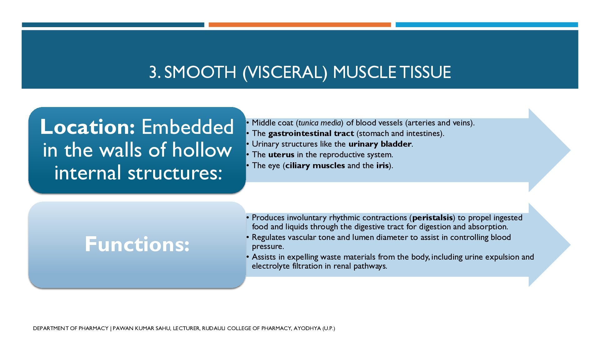
  33. 3. SMOOTH (VISCERAL) MUSCLE TISSUE coat (tunica media) of blood

    vessels (arteries and veins). Location: Embedded •• Middle The gastrointestinal tract (stomach and intestines). • Urinary structures like the urinary bladder. in the walls of hollow • The uterus in the reproductive system. • The eye (ciliary muscles and the iris). internal structures: Functions: • Produces involuntary rhythmic contractions (peristalsis) to propel ingested food and liquids through the digestive tract for digestion and absorption. • Regulates vascular tone and lumen diameter to assist in controlling blood pressure. • Assists in expelling waste materials from the body, including urine expulsion and electrolyte filtration in renal pathways. DEPARTMENT OF PHARMACY | PAWAN KUMAR SAHU, LECTURER, RUDAULI COLLEGE OF PHARMACY, AYODHYA (U.P.)
  34. D) NERVOUS TISSUE Nervous tissue forms the central communication and

    control system of the body. It specializes in receiving sensations, processing information, and sending rapid signals to coordinate body activities. Fundamental Unit: The neuron (nerve cell) is the fundamental structural and functional unit of the nervous system. Neurons do not divide, and they specialize in transmitting electrical signals. DEPARTMENT OF PHARMACY | PAWAN KUMAR SAHU, LECTURER, RUDAULI COLLEGE OF PHARMACY, AYODHYA (U.P.)
  35. STRUCTURAL ANATOMY OF A NEURON A typical neuron consists of

    three main structural regions: the cell body, dendrites, and an axon. DEPARTMENT OF PHARMACY | PAWAN KUMAR SAHU, LECTURER, RUDAULI COLLEGE OF PHARMACY, AYODHYA (U.P.)
  36. 1. CELL BODY (SOMA / CYTON) Structure: It is the

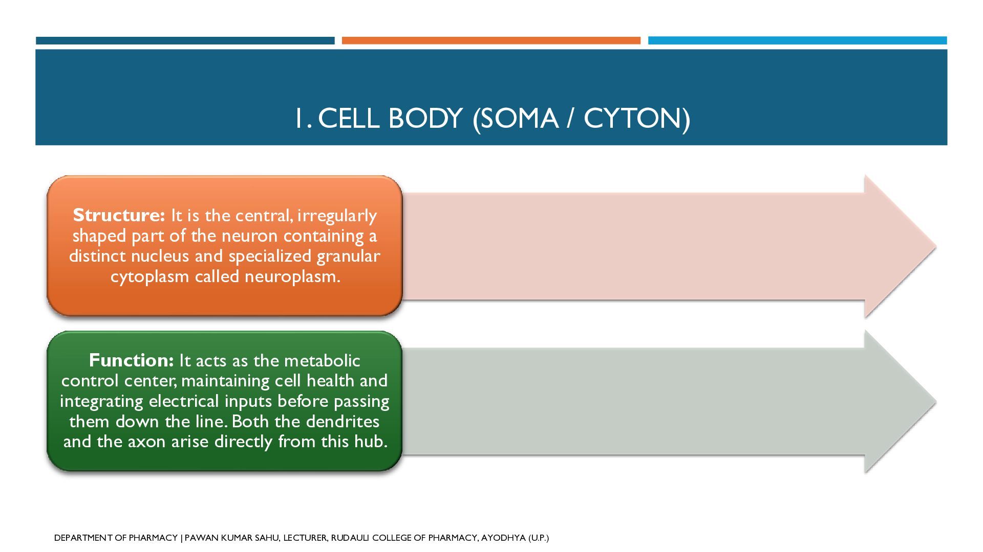
    central, irregularly shaped part of the neuron containing a distinct nucleus and specialized granular cytoplasm called neuroplasm. Function: It acts as the metabolic control center, maintaining cell health and integrating electrical inputs before passing them down the line. Both the dendrites and the axon arise directly from this hub. DEPARTMENT OF PHARMACY | PAWAN KUMAR SAHU, LECTURER, RUDAULI COLLEGE OF PHARMACY, AYODHYA (U.P.)
  37. 2. DENDRITES Structure: These are short, fine, highly branched tree-like

    projections extending outwards from the cell body. Function: They serve as the receiving antennas of the neuron. They collect incoming stimuli from sensory receptors or neighboring neurons and transmit these nerve impulses toward the cell body. DEPARTMENT OF PHARMACY | PAWAN KUMAR SAHU, LECTURER, RUDAULI COLLEGE OF PHARMACY, AYODHYA (U.P.)
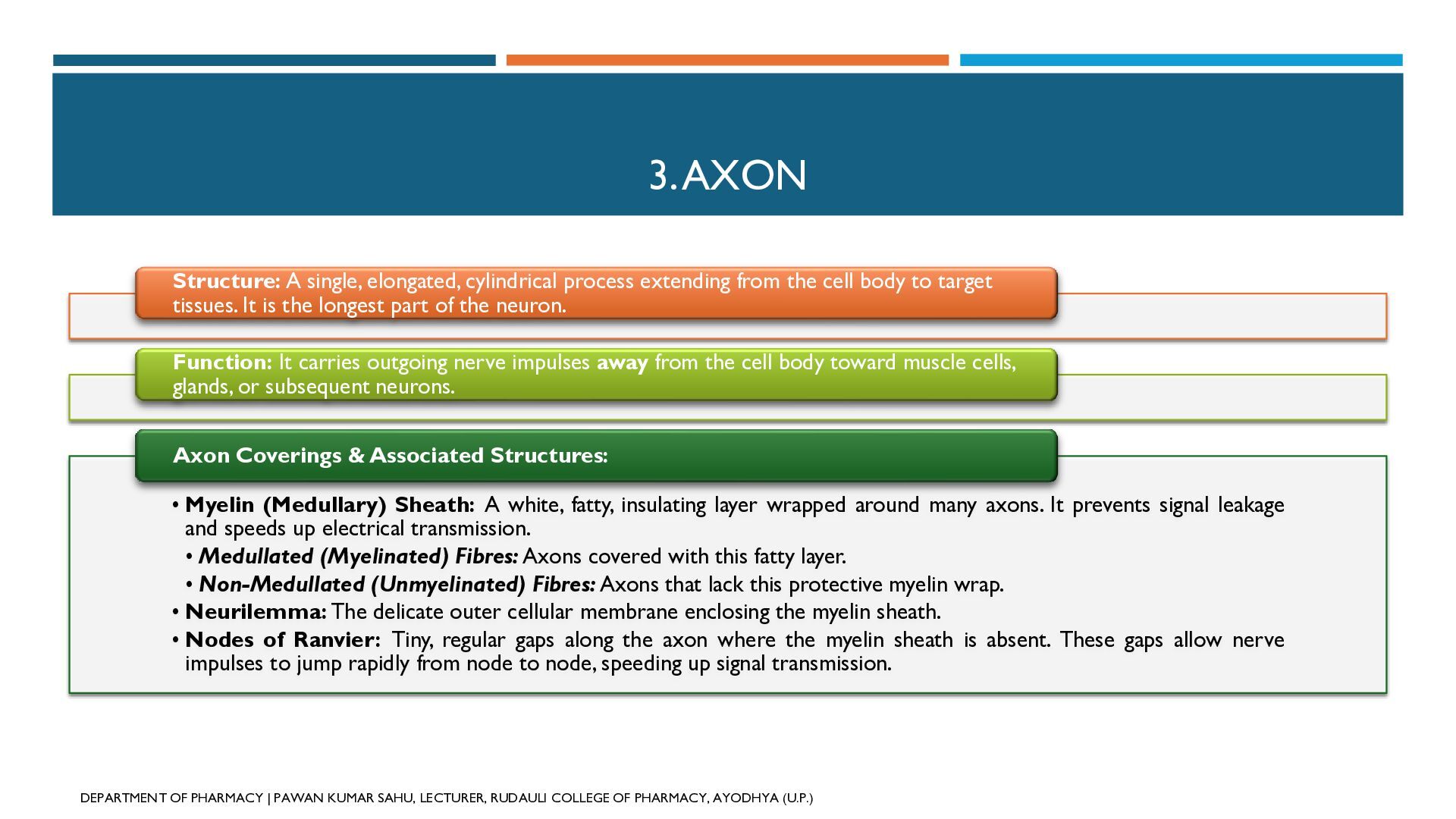
  38. 3. AXON Structure: A single, elongated, cylindrical process extending from

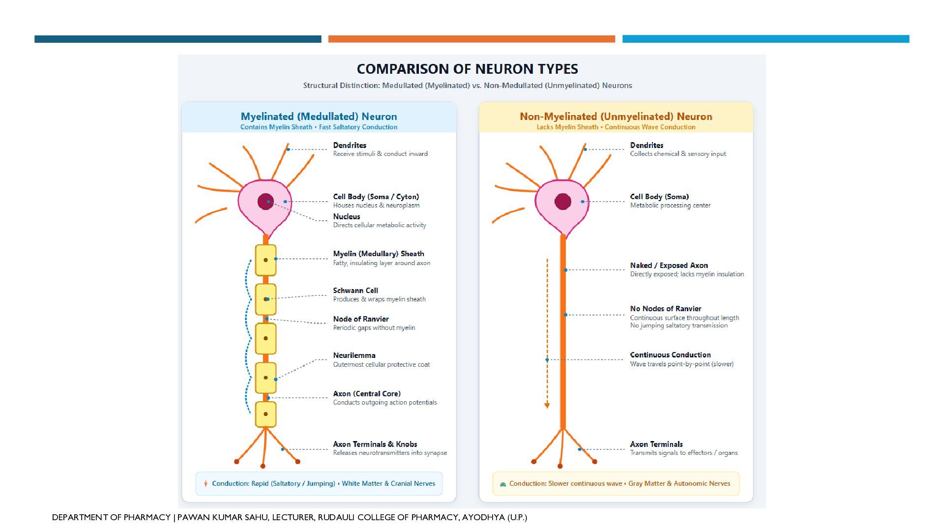
    the cell body to target tissues. It is the longest part of the neuron. Function: It carries outgoing nerve impulses away from the cell body toward muscle cells, glands, or subsequent neurons. Axon Coverings & Associated Structures: • Myelin (Medullary) Sheath: A white, fatty, insulating layer wrapped around many axons. It prevents signal leakage and speeds up electrical transmission. • Medullated (Myelinated) Fibres: Axons covered with this fatty layer. • Non-Medullated (Unmyelinated) Fibres: Axons that lack this protective myelin wrap. • Neurilemma: The delicate outer cellular membrane enclosing the myelin sheath. • Nodes of Ranvier: Tiny, regular gaps along the axon where the myelin sheath is absent. These gaps allow nerve impulses to jump rapidly from node to node, speeding up signal transmission. DEPARTMENT OF PHARMACY | PAWAN KUMAR SAHU, LECTURER, RUDAULI COLLEGE OF PHARMACY, AYODHYA (U.P.)
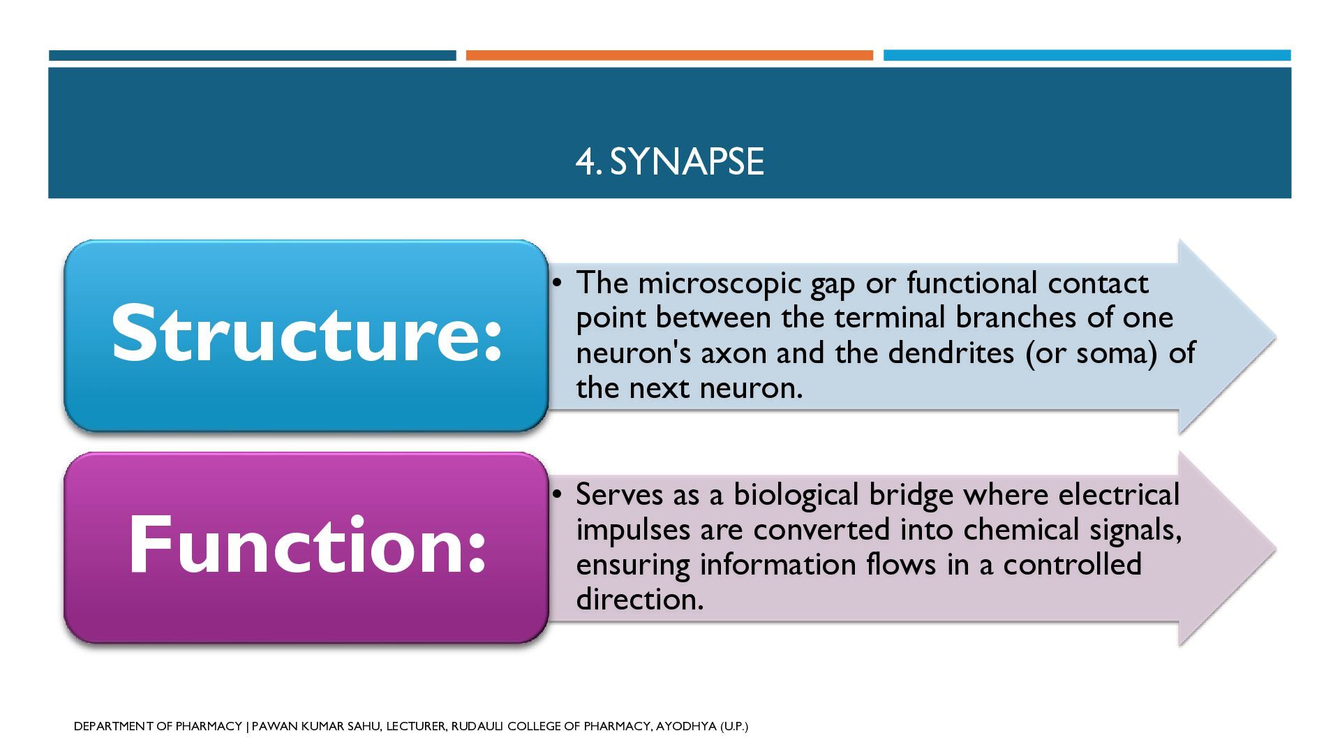
  39. 4. SYNAPSE Structure: • The microscopic gap or functional contact

    point between the terminal branches of one neuron's axon and the dendrites (or soma) of the next neuron. Function: • Serves as a biological bridge where electrical impulses are converted into chemical signals, ensuring information flows in a controlled direction. DEPARTMENT OF PHARMACY | PAWAN KUMAR SAHU, LECTURER, RUDAULI COLLEGE OF PHARMACY, AYODHYA (U.P.)
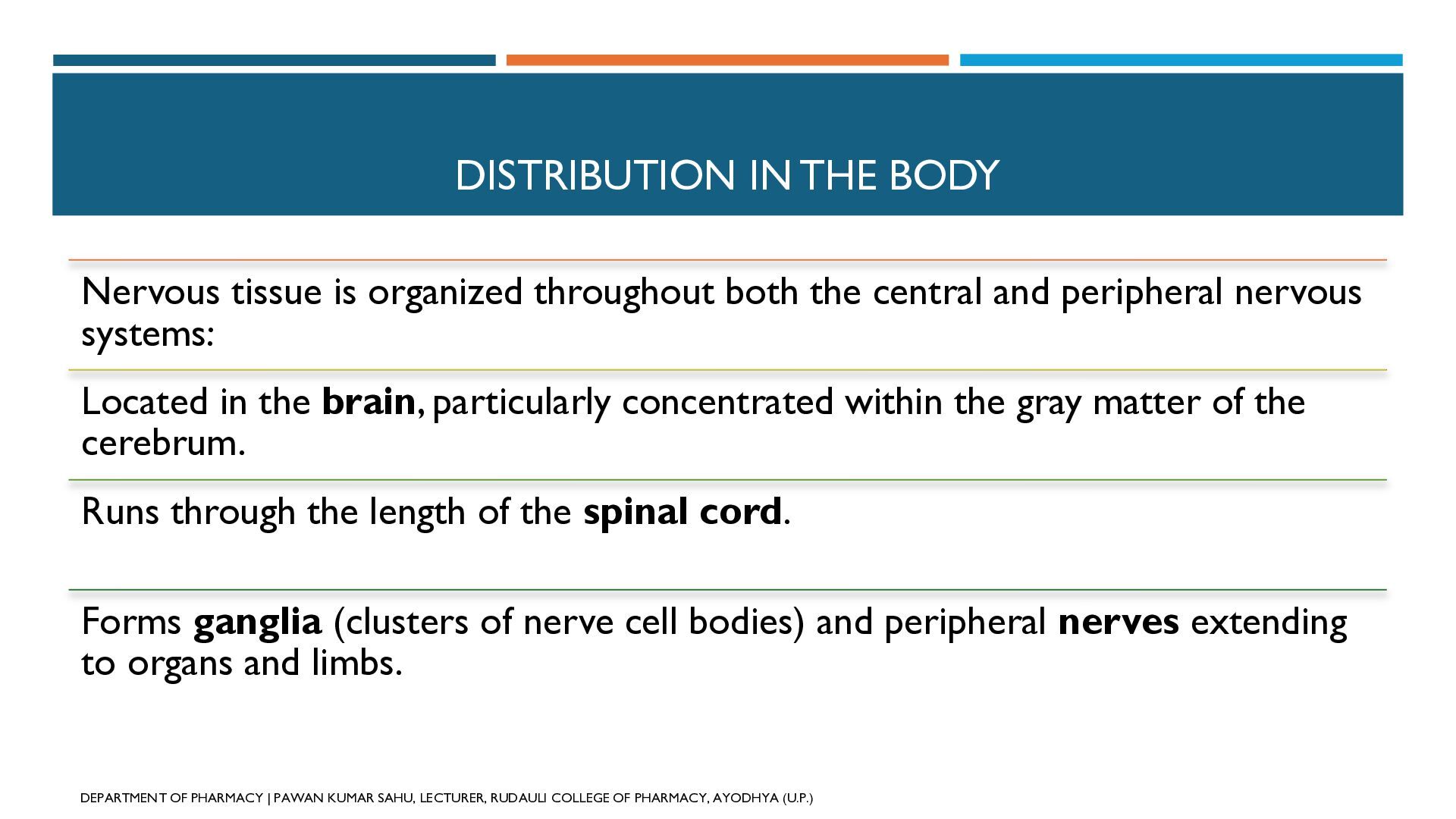
  40. DISTRIBUTION IN THE BODY Nervous tissue is organized throughout both

    the central and peripheral nervous systems: Located in the brain, particularly concentrated within the gray matter of the cerebrum. Runs through the length of the spinal cord. Forms ganglia (clusters of nerve cell bodies) and peripheral nerves extending to organs and limbs. DEPARTMENT OF PHARMACY | PAWAN KUMAR SAHU, LECTURER, RUDAULI COLLEGE OF PHARMACY, AYODHYA (U.P.)
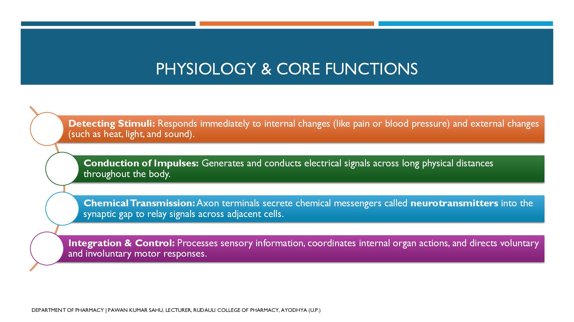
  41. PHYSIOLOGY & CORE FUNCTIONS Detecting Stimuli: Responds immediately to internal

    changes (like pain or blood pressure) and external changes (such as heat, light, and sound). Conduction of Impulses: Generates and conducts electrical signals across long physical distances throughout the body. Chemical Transmission: Axon terminals secrete chemical messengers called neurotransmitters into the synaptic gap to relay signals across adjacent cells. Integration & Control: Processes sensory information, coordinates internal organ actions, and directs voluntary and involuntary motor responses. DEPARTMENT OF PHARMACY | PAWAN KUMAR SAHU, LECTURER, RUDAULI COLLEGE OF PHARMACY, AYODHYA (U.P.)